. 2022 Jan 7;130(1):5-23.
doi: 10.1161/CIRCRESAHA.121.318852. Epub 2021 Nov 18.

VE-Cadherin Is Required for Cardiac Lymphatic Maintenance and Signaling

Affiliations

VE-Cadherin Is Required for Cardiac Lymphatic Maintenance and Signaling

Natalie R Harris et al. Circ Res. .

Abstract

Background: The adherens protein VE-cadherin (vascular endothelial cadherin) has diverse roles in organ-specific lymphatic vessels. However, its physiological role in cardiac lymphatics and its interaction with lymphangiogenic factors has not been fully explored. We sought to determine the spatiotemporal functions of VE-cadherin in cardiac lymphatics and mechanistically elucidate how VE-cadherin loss influences prolymphangiogenic signaling pathways, such as adrenomedullin and VEGF (vascular endothelial growth factor)-C/VEGFR3 (vascular endothelial growth factor receptor 3) signaling.

Methods: Cdh5flox/flox;Prox1CreERT2 mice were used to delete VE-cadherin in lymphatic endothelial cells across life stages, including embryonic, postnatal, and adult. Lymphatic architecture and function was characterized using immunostaining and functional lymphangiography. To evaluate the impact of temporal and functional regression of cardiac lymphatics in Cdh5flox/flox;Prox1CreERT2 mice, left anterior descending artery ligation was performed and cardiac function and repair after myocardial infarction was evaluated by echocardiography and histology. Cellular effects of VE-cadherin deletion on lymphatic signaling pathways were assessed by knockdown of VE-cadherin in cultured lymphatic endothelial cells.

Results: Embryonic deletion of VE-cadherin produced edematous embryos with dilated cardiac lymphatics with significantly altered vessel tip morphology. Postnatal deletion of VE-cadherin caused complete disassembly of cardiac lymphatics. Adult deletion caused a temporal regression of the quiescent epicardial lymphatic network which correlated with significant dermal and cardiac lymphatic dysfunction, as measured by fluorescent and quantum dot lymphangiography, respectively. Surprisingly, despite regression of cardiac lymphatics, Cdh5flox/flox;Prox1CreERT2 mice exhibited preserved cardiac function, both at baseline and following myocardial infarction, compared with control mice. Mechanistically, loss of VE-cadherin leads to aberrant cellular internalization of VEGFR3, precluding the ability of VEGFR3 to be either canonically activated by VEGF-C or noncanonically transactivated by adrenomedullin signaling, impairing downstream processes such as cellular proliferation.

Conclusions: VE-cadherin is an essential scaffolding protein to maintain prolymphangiogenic signaling nodes at the plasma membrane, which are required for the development and adult maintenance of cardiac lymphatics, but not for cardiac function basally or after injury.

Keywords: adrenomedullin; cadherins; endothelial cells; myocardial infarction; vascular endothelial growth factor receptor.


Conflict of interest statement

DISCLOSURES

The authors declare that they have no conflicts of interest.

Figures
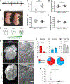
Figure 1:
Figure 1:
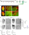
Embryonic Deletion of VE-cadherin Results in Edematous Embryos with Pleural Fluid and Altered Lymphatic Vessel Tip Morphology A) Tamoxifen injection scheme to delete VE-Cadherin in lymphatics during embryogenesis at E14.5 and E15.5. TM, tamoxifen. B) Representative images of Cdh5flox/flox and Prox1CreERT2;Cdh5flox/flox (abbreviated subsequently as Cdh5LEC-KO) embryos at E18.5. Scale bar 0.5 mm. TM administered E13.5 via IP. Scale bar 100 μm. Morphometric and gravimetric analysis of E18.5 embryos: C) body weight, D) crown-rump length, E) heart weight, F) ratio of heart weight:crown rump length. Data displayed as mean ± SD (n = 9 for Cdh5flox/flox, n = 8 for Cdh5LEC-KO). G) Cartoon depicting embryonic cardiac lymphatic development E13.5-E18.5. H) Embryonic hearts at E18.5 stained with LYVE1 (white). Scale bar 100μm. Dashed white box represents inset location. LYVE1 vessel tips in “Inset” with blunt morphology marked by blue arrows and tips with spiky morphology marked by red arrows. Scale bar 20μm. Quantification of LYVE1 vessel tip morphology: I) number of spiky tips, J) Number of blunt tips, K) total number of tips. Data displayed mean ± SD (n = 4 for Cdh5flox/flox, n = 4 for Cdh5LEC-KO). L) Pie chart depicting average percentage of blunt vs. spiky tips for Cdh5flox/flox and Cdh5LEC-KO. M) Ratio of spiky tips to blunt tips Data displayed mean ± SD (n = 4 for Cdh5flox/flox, n = 4 for Cdh5LEC-KO). An unpaired Student’s t-test with Welch’s correction was used to compare between genotypes (C-F). Mann-Whitney Test was used to compare between genotypes (I-K, and M). *p<0.05 and **p<0.01. Detailed description of group size, normality testing, statistical tests and post-tests, and exact p-values can be found in the statistical supplement.
Figure 2:
Figure 2:. Postnatal Deletion of VE-cadherin Leads to Loss of Cardiac Lymphatic Network
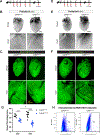
A) Tamoxifen injection scheme to delete VE-Cadherin in lymphatics postnatally at P1 and P3, with analysis at indicated time points. B) Cartoon depicting cardiac lymphatic development E18.5-P15. C) P31 Hearts whole-mount stained for SMA imaged for SMA and Prox1-GFP. Scale bar 200 μm. Arrows point to isolated LECs in the Cdh5LEC-KO hearts. D) Adult (3 months) hearts whole mount stained for podoplanin to confirm absence of cardiac lymphatic is maintained into adulthood. Scale bar 500 μm. E) Quantification of vessel percentage area in both Cdh5flox/flox and Cdh5LEC-KO hearts. Data displayed at Individual points are the averaged vessel percentage area of both the dorsal and ventral sides of each heart. (n=6 Cdh5flox/flox, n=3 Cdh5LEC-KO from two litters). F) Echocardiography measurements displaying no statistical difference in function between genotypes, and a trend towards smaller left ventricular (LV) mass, p = 0.0635. Data displayed as mean ± SD (n=4 Cdh5flox/flox, n=5 Cdh5LEC-KO from two litters). A Mann Whitney test was used to compare between genotypes (E-F). *p<0.05. Detailed description of group size, normality testing, statistical tests and post-tests, and exact p-values can be found in the statistical supplement.
Figure 3:
Figure 3:. Loss of VE-cadherin in Adults Leads to Progressive Regression of Cardiac Lymphatics, Compatible with Long-Term Survival
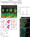
A) Tamoxifen injection scheme to delete VE-cadherin in lymphatics in adult mice, with analysis at day 22. B) Hearts whole-mount stained with podoplanin. Scale bar 200 μm. C) Hearts fluorescence imaged for Prox1-GFP. Yellow brackets on inset indicate discontinuous Prox1-GFP positive vessels and punctate islands of Prox1-GFP positive LECs. Scale bar 100 μm. D) Tamoxifen injection scheme to delete VE-cadherin in lymphatics in adult mice, with analysis at day 43. E) Hearts whole-mount stained with podoplanin. Scale bar = 200 μm, Scale bar = 80 μm for inset. F) Hearts fluorescence imaged for Prox1-GFP. Yellow brackets on inset indicate islands of Prox1-GFP positive LEC islands, which are reduced in number in comparison to D22. Scale bar = 100 μm, Scale bar = 60 μm for inset. G) Angiotool quantification of vessel percentage area in both Cdh5flox/flox and Cdh5LEC-KO at both the D22 and D43 analysis timepoints (Cdh5flox/flox: D22|n=8, D43|n=8 ; Cdh5LEC-KO: D22|n=10, D43|n=11). All values are mean ± SD. H) Characterization of the Prox1-GFP+ population of non-myocyte cells by flow cytometry. Hearts were collected from tamoxifen treated Cdh5flox/flox and Cdh5LEC-KO at D43, mechanically and enzymatically digested (n=1 per genotype). Cell suspension was run through a 40-um filter to remove cardiomyocytes and remaining cell suspension was labeled with Hoechst, to mark live cells, and analyzed by FACS. Flow cytometry dot plots for GFP displayed; cell populations expressed as percentages (%) of scatter/singlets from digested cardiac tissue. SCC, side scatter; BL1-A::Prox1-GFP-GFP; indicates filter selected for GFP signal. A two-way ANOVA with a Tukey’s multiple comparisons post-test was used to compare vessel percentage area between genotypes and timepoints. **p<0.01, ***p<0.001, and ****p<0.0001. Detailed description of group size, normality testing, statistical tests and post-tests, and exact p-values can be found in the statistical supplement.
Figure 4:
Figure 4:. Loss of VE-cadherin leads to basal cardiac lymphatic dysfunction, results in increased infarct size and blunts cardiac lymphangiogenesis after myocardial infarction (MI).
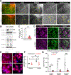
A) Tamoxifen injection scheme to delete VE-cadherin in lymphatics in adult mice, indicating assessment of basal cardiac lymphatic function analysis at day 22, Pre-MI assessment of basal cardiac function by conscious echocardiography and induction of MI by permanent ligation of the left anterior descending artery (LAD) performed at D43. Assessment of Post-MI cardiac function performed routinely between D43 and D63 (Table 2). Hearts collected 20 days-post MI (D63) and histological analysis of the heart and evaluation of lymphatic coverage was performed. B) Cardiac lymphangiography of Cdh5flox/flox and Cdh5LEC-KO. Fluorescent quantum dots (Qdot 605, red) are injected intramyocardially in the apex of the heart and selectively taken up by Prox1-GFP (green) superficial cardiac lymphatics and transported towards draining lymph nodes of the heart. Yellow signal indicated Prox1-GFP+ vessels containing Qdot605. Leakage of Qdot from Prox1-GFP+ lymphatics denoted by white bars. Scale bar 100 μm, Scale bar = 60 μm for inset. C) Gravimetric analysis of cardiac water content in uninjured Cdh5flox/flox and Cdh5LEC-KO at D117. Portion of heart collected, weighed and dried for 6 days at 65°C. Ratio wet weight to dry weight displayed (n = 4 for Cdh5flox/flox and n = 4 for Cdh5LEC-KO). D) Histological Analysis of cardiac sections 20 days post-MI. Left: Hematoxylin-eosin (H&E), Middle: Masson’s Trichrome, Right: Sirus Red. Scale bar = 500 μm for all three staining sets. E) Quantification of Infarct Area using Sirius Red Staining Intensity (n = 6 for Cdh5flox/flox and n = 5 for Cdh5LEC-KO). All values are mean ± SD. F) Immunohistochemistry of cardiac sections in infarcted Cdh5flox/flox and Cdh5LEC-KO 20 days post-MI (yellow=LYVE1, Magenta=Podoplanin, green=Prox1-GFP, blue=DAPI. Podoplanin positive epicardium denoted with white arrow. Scale bar 1000 μm. A Mann Whitney test was used to compare between genotypes. **p<0.01. Detailed description of group size, normality testing, statistical tests and post-tests, and exact p-values can be found in the statistical supplement.
Figure 5:
Figure 5:. Loss of VE-cadherin Results in Internalization of VEGFR3 and Attenuated VEGFR3 Signaling
A) Hearts whole-mount stained with LYVE1 and VEGFR3. Yellow boxed areas indicate the zoomed in area displayed in the inset panels. Yellow arrows indicate cardiac lymphatics in which VEGFR3 signal is decreased or absent from the indicated lymphatic vessel in the Cdh5LEC-KO heart. Scale bar = 500 μm, Scale bar = 150 μm for inset. B) Representative western blot analysis of CDH5 and VEGFR3 levels after CDH5 knockdown. C) Quantification of CDH5 knockdown normalized to CDH5 levels in shScramble treated control group from n=3 independent experiments. D) Representative images of VE-cadherin and VEGFR3 cellular localization in confluent LECs which were treated with shScramble or shCDH5. Scale bar 20 μm. E) Representative images of VEGFR3/p-Tyr PLA generated foci in shScramble or shCDH5 treated lymphatic endothelial cells (LECs) which were treated with DMSO or VEGF-C to induce VEGF-C-mediated VEGFR3 phosphorylation. Scale bar 20 μm. F) Quantification of the number of foci per cell. Each point represents average of an experiment, n = 4 independent experiments. G) Proliferation in response to VEGFC stimulation after CDH5 knockdown. Data shown as percentage of Edu positive cells (n = 6 independent experiments for shScramble, n = 5 independent experiments for shCDH5). Each point represents average of an individual experiment. All values are mean ± SD. An unpaired Student’s t-test with Welch’s correction was used to compare between treatment groups. Significance for multiple comparisons was determined by two-way ANOVA with Sidak multiple comparisons test. *p<0.05, **p<0.01, ****p<0.0001. Detailed description of group size, normality testing, statistical tests and post-tests, and exact p-values can be found in the statistical supplement.
Figure 6:
Figure 6:. Adrenomedullin signaling transactivates VEGFR3 in Lymphatic Endothelial Cells
A) Representative images of VEGFR3/p-Tyr PLA generated foci in treated lymphatic endothelial cells (LECs). Scale bar 20 μm. (B) Quantification of the number of foci per cell (n=5 independent experiments). In all graphs the red horizontal line represents the mean. Significance for multiple comparisons was determined by one-way ANOVA with Bonferroni posttests. (C) VEGFR3 immunoprecipitated from treated LECs was blotted for VEGFR3 and tyrosine phosphorylation. D) Representative images of VEGFR3/p-Tyr PLA generated foci in shScramble or shCDH5 treated LECs then subject to a pretreatment of sunitinib before administration of DMSO, AM, or AM-22–52, used to assess VEGFR3 transactivation (AM 22–52 alone and VEGF-C images not shown). E) Quantification of number of foci per cell (n=4 independent experiments). Significance for multiple comparisons was determined by two-way ANOVA with Tukey’s multiple comparisons test. All values are mean ± SD.*p<0.05 and **p<0.01. Detailed description of group size, normality testing, statistical tests and post-tests, and exact p-values can be found in the statistical supplement.
Figure 7:
Figure 7:. VE-cadherin is a Critical Component of the VEGFR3 Signaling Node within LECs responsible for Cardiac Lymphatic Maintenance and Function
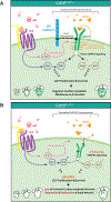
A) Schematic depicting the role of VE-cadherin in stabilizing VEGFR3 at the membrane in LECs allowing for VEGFC ligand-mediated activation of VEGFR3 or adrenomedullin mediated transactivation of VEGFR3 through binding of AM to CLR/RAMP complex triggering c-Src mediated phosphorylation of VEGFR3. This stabilized VEGFR3 signaling node in the LECs of Cdh5flox/flox mice promotes LEC proliferation and survival, allowing for normal maintenance and function of the cardiac lymphatic network. B) Loss of VE-cadherin leads to a destabilized VEGFR3 signaling node, attenuating downstream signaling in LECs, which in Cdh5EC-KO mice ultimately leads to a failure to form a proper cardiac lymphatic network when VE-cadherin is lost postnatally. When VE-cadherin is lost in adulthood, the fully developed cardiac lymphatic network regresses and leads to basal lymphatic dysfunction.

Comment in

References

    1. Carmeliet P, Lampugnani MG, Moons L, Breviario F, Compernolle V, Bono F, Balconi G, Spagnuolo R, Oosthuyse B, Dewerchin M, Zanetti A, Angellilo A, Mattot V, Nuyens D, Lutgens E, Clotman F, de Ruiter MC, Gittenberger-de Groot A, Poelmann R, Lupu F, Herbert JM, Collen D, Dejana E. Targeted deficiency or cytosolic truncation of the ve-cadherin gene in mice impairs vegf-mediated endothelial survival and angiogenesis. Cell 1999;98:147–157 - PubMed
    1. Corada M, Mariotti M, Thurston G, Smith K, Kunkel R, Brockhaus M, Lampugnani MG, Martin-Padura I, Stoppacciaro A, Ruco L, McDonald DM, Ward PA, Dejana E. Vascular endothelial-cadherin is an important determinant of microvascular integrity in vivo. Proc Natl Acad Sci U S A 1999;96:9815–9820 - PMC - PubMed
    1. Lagendijk AK, Hogan BM. Ve-cadherin in vascular development: A coordinator of cell signaling and tissue morphogenesis. Curr Top Dev Biol 2015;112:325–352 - PubMed
    1. Baluk P, Fuxe J, Hashizume H, Romano T, Lashnits E, Butz S, Vestweber D, Corada M, Molendini C, Dejana E, McDonald DM. Functionally specialized junctions between endothelial cells of lymphatic vessels. J Exp Med 2007;204:2349–2362 - PMC - PubMed
    1. Yang Y, Cha B, Motawe ZY, Srinivasan RS, Scallan JP. Ve-cadherin is required for lymphatic valve formation and maintenance. Cell Rep 2019;28:2397–2412 e2394 - PMC - PubMed

Publication types