. 2021 Nov 17;21(1):607.
doi: 10.1186/s12935-021-02306-y.

Anticancer effects of mifepristone on human uveal melanoma cells

Affiliations

Anticancer effects of mifepristone on human uveal melanoma cells

Prisca Bustamante Alvarez et al. Cancer Cell Int. .

Abstract

Background: Uveal melanoma (UM), the most prevalent intraocular tumor in adults, is a highly metastatic and drug resistant lesion. Recent studies have demonstrated cytotoxic and anti-metastatic effects of the antiprogestin and antiglucocorticoid mifepristone (MF) in vitro and in clinical trials involving meningioma, colon, breast, and ovarian cancers. Drug repurposing is a cost-effective approach to bring approved drugs with good safety profiles to the clinic. This current study assessed the cytotoxic effects of MF in human UM cell lines of different genetic backgrounds.

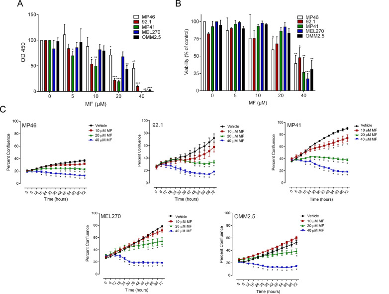
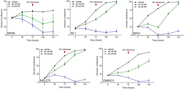
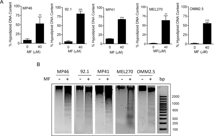
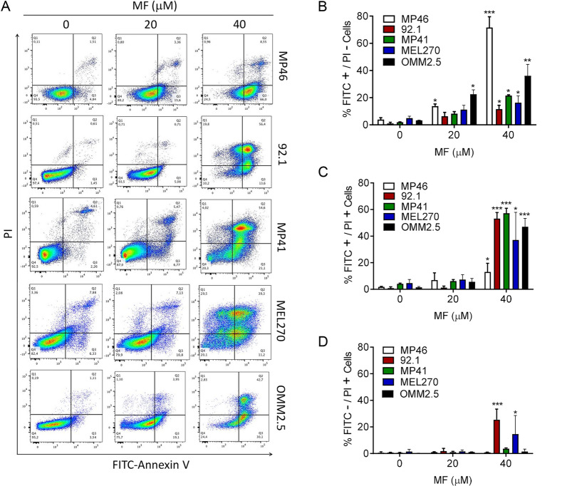
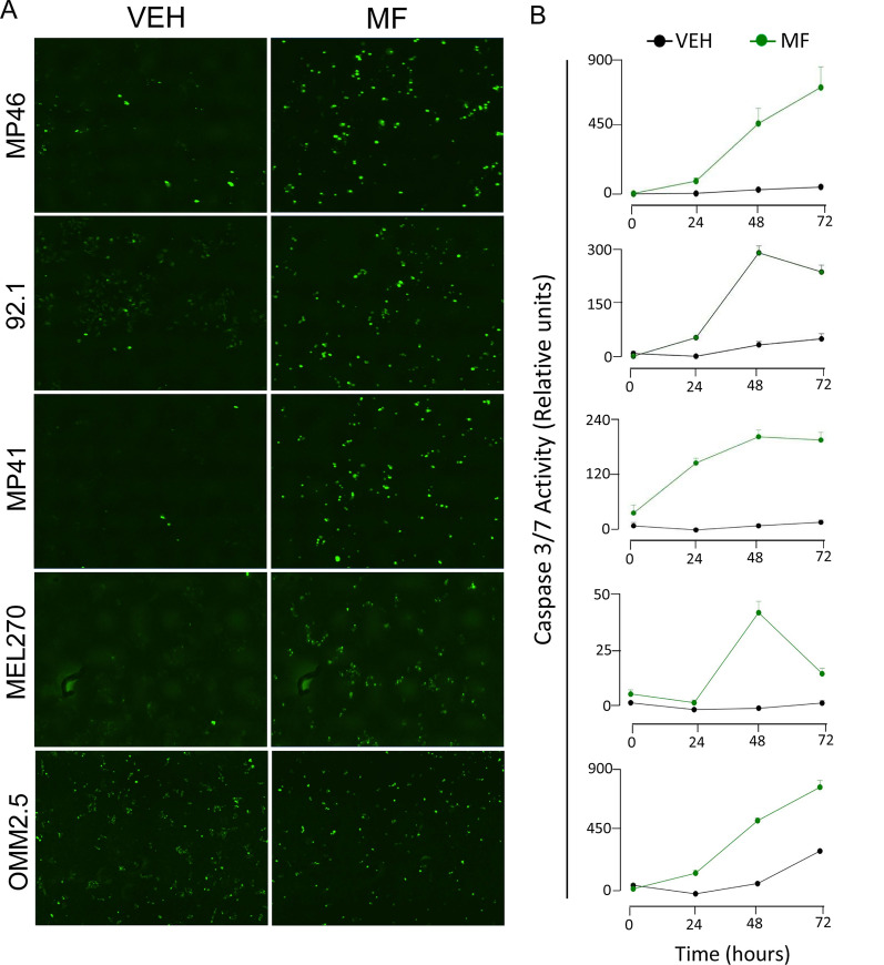
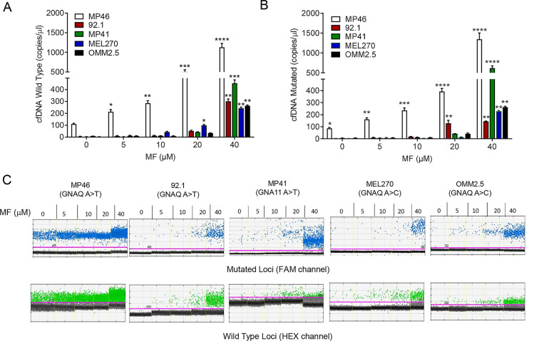
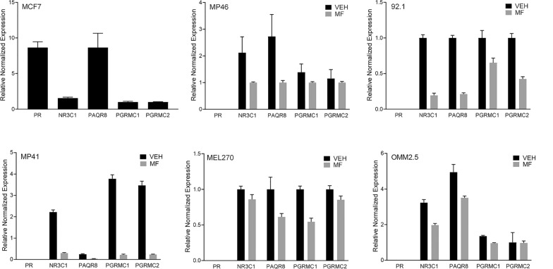
Methods: The effects of incremental concentrations of MF (0, 5, 10, 20, or 40 μM) on a panel of human UM primary (MEL270, 92.1, MP41, and MP46) and metastatic (OMM2.5) cells were evaluated. Cells were incubated with MF for up to 72 h before subsequent assays were conducted. Cellular functionality and viability were assessed by Cell Counting Kit-8, trypan blue exclusion assay, and quantitative label-free IncuCyte live-cell analysis. Cell death was analyzed by binding of Annexin V-FITC and/or PI, caspase-3/7 activity, and DNA fragmentation. Additionally, the release of cell-free DNA was assessed by droplet digital PCR, while the expression of progesterone and glucocorticoid receptors was determined by quantitative real-time reverse transcriptase PCR.

Results: MF treatment reduced cellular proliferation and viability of all UM cell lines studied in a concentration-dependent manner. A reduction in cell growth was observed at lower concentrations of MF, with evidence of cell death at higher concentrations. A significant increase in Annexin V-FITC and PI double positive cells, caspase-3/7 activity, DNA fragmentation, and cell-free DNA release suggests potent cytotoxicity of MF. None of the tested human UM cells expressed the classical progesterone receptor in the absence or presence of MF treatment, suggesting a mechanism independent of the modulation of the cognate nuclear progesterone receptor. In turn, all cells expressed non-classical progesterone receptors and the glucocorticoid receptor.

Conclusion: This study demonstrates that MF impedes the proliferation of UM cells in a concentration-dependent manner. We report that MF treatment at lower concentrations results in cell growth arrest, while increasing the concentration leads to lethality. MF, which has a good safety profile, could be a reliable adjuvant of a repurposing therapy against UM.

Keywords: Cancer therapy; Drug repurposing; Mifepristone; Uveal melanoma.


Conflict of interest statement

The authors declare that they have no competing interests.

Figures

Fig. 1
Fig. 1
MF inhibits functionality, growth capacity, and viability of UM cell lines in a concentration-related manner. Graphs represent the level of cellular functionality or viability, respectively as detected via a CCK8 colorimetric assay (A) or Trypan Blue exclusion assay (B) after cells were treated with increasing concentrations of MF (0, 5, 10, 20, or 40 μM) for 72 h. C Growth curves obtained through Incucyte live cell imaging system, tracking cellular confluency. In A and B, data were analyzed using two-way ANOVA followed by Dunnett’s multiple comparison test. In C, data were analyzed using repeated measures ANOVA followed by Tuckey’s multiple comparison test.* Indicates p < 0.05, ** indicates p < 0.01, whereas *** indicates p < 0.001 compared against vehicle-treated controls
Fig. 2
Fig. 2
Long-term toxicity of MF towards UM cell lines and the consequence of MF withdrawal. UM cells were treated with MF at concentrations of either 0, 20, or 30 μM for 72 h and imaged every 6 h in the Incucyte. Following the initial 72 h, media was aspirated, replaced with regular growth media, and placed back into the Incucyte to be imaged for another 72 h. The red arrows at 72 h indicate the moment in which MF was removed from the media. Data were analyzed using repeated measures ANOVA followed by Tuckey’s multiple comparison test. * Indicates p < 0.05, ** indicates p < 0.01, whereas *** indicates p < 0.001 compared against vehicle-treated controls
Fig. 3
Fig. 3
MF induces accumulation of hypodiploid DNA content and DNA ladder. A Quantification of particles with hypodiploid DNA content upon 72 h of MF treatment in a panel of UM cell lines. The hypodiploid DNA content corresponds to the Sub-G1 DNA content extrapolated when performing the cell cycle analysis of the cells treated with MF (the quantitative details are shown in the green-stained sections of the histograms in Additional file 3: Fig. S3). B A similar experiment was done in which all floating and adherent cells were pelleted, gDNA isolated, subjected to agarose electrophoresis, stained with SYBR Gold nuclei acid stain, and imaged. A 100 base pair (bp) maker was run in parallel. −: vehicle; + : 40 µM MF
Fig.4
Fig.4
MF induces apoptosis in UM cells. A Representative histograms depicting the distribution of UM cells exposed to vehicle, 20, or 40 µM MF, and stained with Annexin V-FITC and/or PI after 72 h of incubation. The histograms represent flow cytometry data. B The bar graphs depict the percent of UM cells undergoing early apoptosis as marked by the labeling with only Annexin V-FITC. C Results show the percent of UM cells undergoing late apoptosis represented by cells double labeled with Annexin V-FITC and PI. D The percent of cells likely undergoing necrosis is shown as PI only stained cells. Data were analyzed using two-way ANOVA followed by Dunnett’s multiple comparison test. * Indicates p < 0.05, ** indicates p < 0.01, whereas *** indicates p < 0.001 compared against vehicle-treated controls
Fig. 5
Fig. 5
MF-associated UM cell death is related with the activation of executer caspase-3/7. A Green nuclear staining is generated upon a chemical reaction catalyzed by either active caspases 3 or 7. The images shown represent the endpoint of an experiment done for 72 h following MF treatment at a 40 µM concentration. These images can be observed over imposed with phase contrast in Additional file 5: Fig. S5. B Depicted are the time-course quantifications of the green fluorescence expressed as relative activity with respect to the fluorescence generated by vehicle-treated cells
Fig. 6
Fig. 6
MF treatment induces the release of cell-free DNA into the media supernatant. Graphs show number of wild type (A) and mutant (B) copies of cfDNA per μl of cell-free media obtained 72 h after incubation with vehicle, 5, 10, 20, or 40 µM MF. C Representative one-dimensional plot of mutant GNAQ/GNA11 or wild type cfDNA extracted from conditioned media after treatment for 72 h with the depicted concentrations of MF. Channel compatible with FAM dye shows droplets with mutant target in blue. Wild type target is shown in green using a HEX label. Threshold (pink line) set in between positive (mutant or wild type) and no DNA target (black) droplets
Fig. 7
Fig. 7
The effect of MF in UM cells is independent from the classical nuclear progesterone receptor. SybrGreen-based Real Time PCR quantified the gene expression profiles of PR, PAQR8, PGRMC1, PGRMC2, and NR3C1. β-Actin was used as a reference gene. mRNAs were amplified from either untreated cells or cells treated with 20 µM MF. The mRNA from MCF-7 cells was used as a positive control for the expression of the classical PR. No template control and no reverse transcriptase control were added in each assay. Individual runs were performed in triplicates

References

    1. Chang AEKL, Menck HR. The National Cancer Data Base report on cutaneous and noncutaneous melanoma: a summary of 84,836 cases from the past decade. The American College of Surgeons Commission on Cancer and the American Cancer Society. Cancer. 1998;83(8):1664–1678. - PubMed
    1. Ghazawi FM, Darwich R, Le M, Rahme E, Zubarev A, Moreau L, Burnier JV, Sasseville D, Burnier MN, Litvinov IV. Uveal melanoma incidence trends in Canada: a national comprehensive population-based study. Br J Ophthalmol. 2019;103(12):1872–1876. - PubMed
    1. Rozeman EA, Dekker TJA, Haanen J, Blank CU. Advanced melanoma: current treatment options, biomarkers, and future perspectives. Am J Clin Dermatol. 2018;19(3):303–317. - PubMed
    1. van der Kooij MK, Speetjens FM, van der Burg SH, Kapiteijn E. Uveal versus cutaneous melanoma; same origin, very distinct tumor types. Cancers (Basel) 2019;11(6):845. - PMC - PubMed
    1. Yang J, Manson DK, Marr BP, Carvajal RD. Treatment of uveal melanoma: where are we now? Ther Adv Med Oncol. 2018;10:1758834018757175. - PMC - PubMed

Grants and funding