. 2021 Nov 17;19(1):374.
doi: 10.1186/s12951-021-01119-5.

Macrophage membrane camouflaged reactive oxygen species responsive nanomedicine for efficiently inhibiting the vascular intimal hyperplasia

Affiliations

Macrophage membrane camouflaged reactive oxygen species responsive nanomedicine for efficiently inhibiting the vascular intimal hyperplasia

Boyan Liu et al. J Nanobiotechnology. .

Abstract

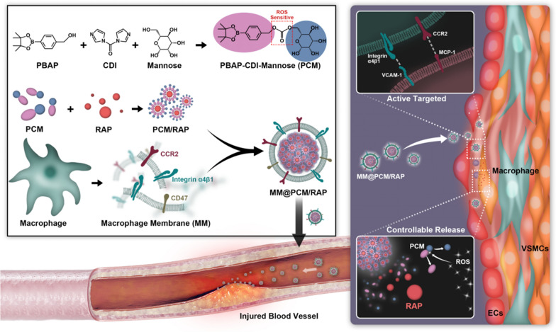
Background: Intimal hyperplasia caused by vascular injury is an important pathological process of many vascular diseases, especially occlusive vascular disease. In recent years, Nano-drug delivery system has attracted a wide attention as a novel treatment strategy, but there are still some challenges such as high clearance rate and insufficient targeting.

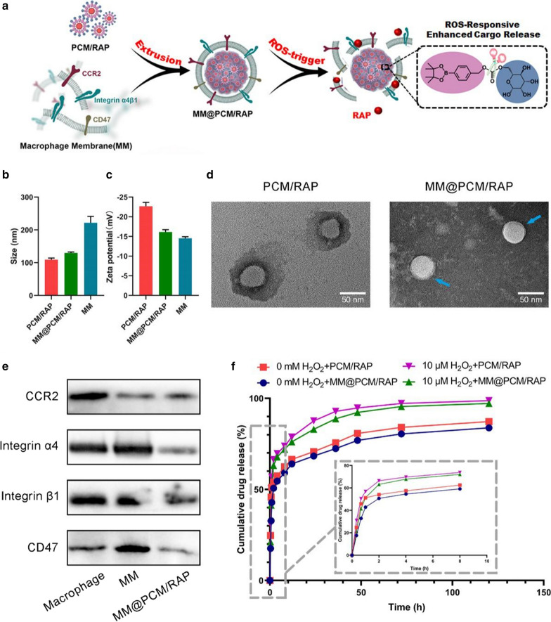
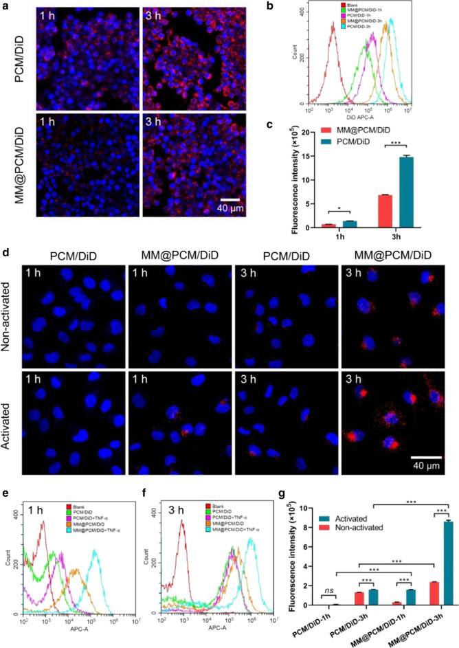
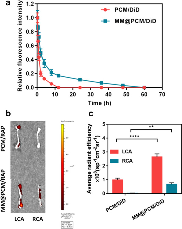
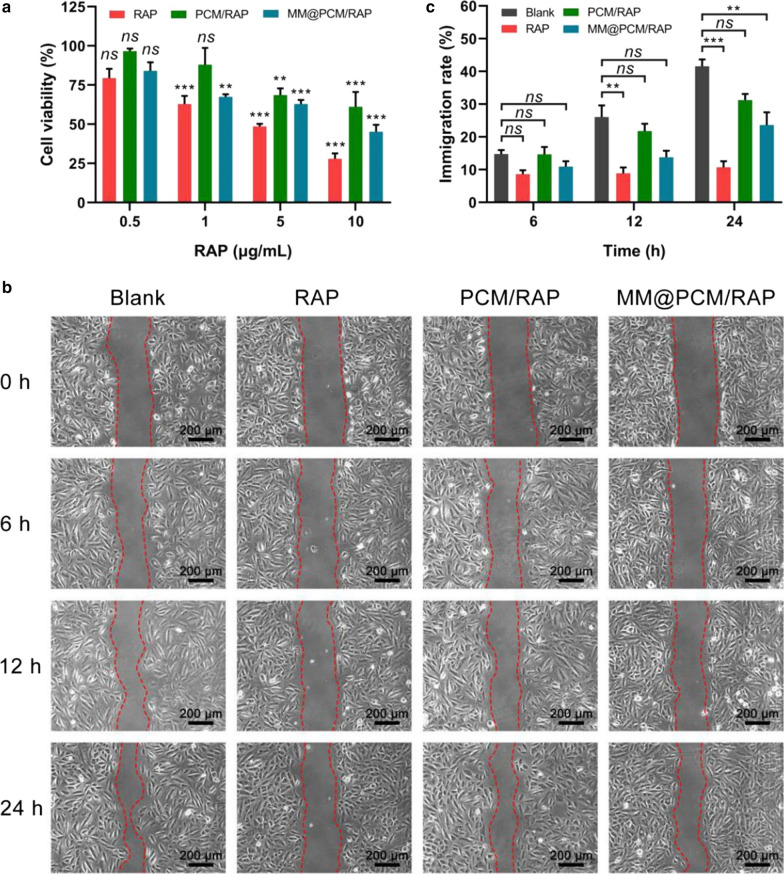
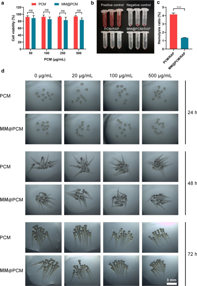
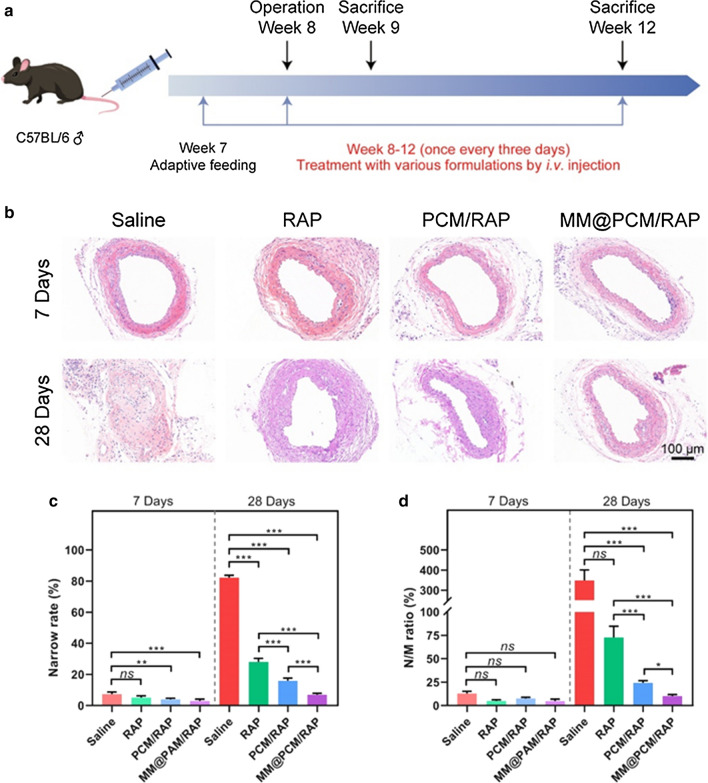
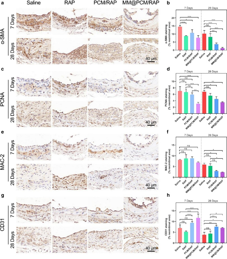
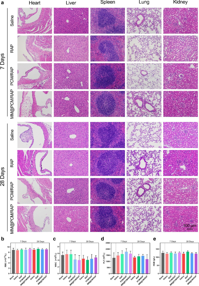
Results: In this study, we report a biomimetic ROS-responsive MM@PCM/RAP nanoparticle coated with macrophage membrane. The macrophage membrane with the innate "homing" capacity can superiorly regulate the recruitment of MM@PCM/RAP to inflammatory lesion to enhance target efficacy, and can also disguise MM@PCM/RAP nanoparticle as the autologous cell to avoid clearance by the immune system. In addition, MM@PCM/RAP can effectively improve the solubility of rapamycin and respond to the high concentration level of ROS accumulated in pathological lesion for controlling local cargo release, thereby increasing drug availability and reducing toxic side effects.

Conclusions: Our findings validate that the rational design, biomimetic nanoparticles MM@PCM/RAP, can effectively inhibit the pathological process of intimal injury with excellent biocompatibility.

Keywords: Intimal hyperplasia; Macrophages; Nanomedicine; ROS-responsive; Targeted delivery.


Conflict of interest statement

The authors declare no conflict of interest.

Figures

Fig. 1
Fig. 1
Illustrations of MM@PCM/RAP for the treatment of IH
Fig. 2
Fig. 2
Characterization of PCM/RAP and MM@PCM/RAP. a Schematic illustration of preparation and drug release of MM@PCM/RAP. b The hydrated particle size and c zeta potential of PCM/RAP, MM@PCM/RAP and MM (n = 3, mean ± SD). d TEM images of PCM/RAP and MM@PCM/RAP (scale bar = 50 nm). e Western blot analysis of CCR2, Integrin α4, Integrin β1 and CD47 in Macrophage, MM and MM@PCM/RAP. f In vitro drug release study of PCM/RAP and MM@PCM/RAP without and with 10 μM H2O2 (n = 3, mean ± SD)
Fig. 3
Fig. 3
In vitro cellular uptake of PCM/DiD and MM@PCM/DiD by RAW 264.7 macrophages and ECs. Nuclei were stained with DAPI (blue), whereas nanoparticles were stained with DiD (red). a CLSM images of PCM/DiD and MM@PCM/DiD phagocytosed by RAW 264.7 cells at 1 h or 3 h (scale bar = 40 µm). b FACS images and c quantification anilysis of PCM/DiD and MM@PCM/DiD phagocytosed by RAW 264.7 cells at 1 h or 3 h (n = 3, mean ± SD). d CLSM images of PCM/DiD and MM@PCM/DiD cellular uptake by ECs without or with TNF-α activated at 1 h or 3 h (scale bar = 40 µm). e, f FACS images of PCM/DiD and MM@PCM/DiD cellular uptake by ECs without or with TNF-α activated at 1 h or 3 h. g Quantification of cellular uptake of PCM/DiD and MM@PCM/DiD in ECs without or with TNF-α activated at 1 h or 3 h (n = 3, mean ± SD). Significances among the groups were determined by one-way ANOVA, followed by post hoc pairwise comparisons with the Tukey or Dunn honest significant difference. *p < 0.05, **p < 0.01, and ***p < 0.001. ns no significance
Fig. 4
Fig. 4
a Pharmacokinetic studies of MM@PCM/DiD and PCM/DiD, and b the ex vivo fluorescence image and c quantitative data of carotid artery (LCA: left carotid artery; RCA: right carotid artery) in mice (n = 5). **p < 0.01, and ****p < 0.0001
Fig. 5
Fig. 5
In vitro VSMCs inhibition. a Viability of VSMCs treated with free RAP, PCM/RAP, and MM@PCM/RAP, respectively, at 0.5, 1, 5 and 10 μg/mL RAP (n = 5, mean ± SD). b Photographs and c quantification analysis of VSMCs migration treated with free RAP, PCM/RAP, and MM@PCM/RAP, respectively, at 2.5 μg/mL RAP (scale bar = 200 µm, n = 5, mean ± SD). Significances among the groups were determined by one-way ANOVA, followed by post hoc pairwise comparisons with the Tukey or Dunn honest significant difference. **p < 0.01, and ***p < 0.001. ns, no significance
Fig. 6
Fig. 6
In vitro biosafety evaluation. a Endothelial toxicity of carrier PCM and MM@PCM (n = 5, mean ± SD). b The images and c quantification analysis of the hemolysis assay of PCM/RAP and MM@PCM/RAP (n = 3, mean ± SD). d Toxic effects of different concentrations of PCM and MM@PCM on zebrafish embryos (scale bar = 3 mm). Significances among the groups were determined by one-way ANOVA, followed by post hoc pairwise comparisons with the Tukey or Dunn honest significant difference. ***p < 0.001. ns no significance
Fig. 7
Fig. 7
Therapeutic effects of i.v.-delivered MM@PCM/RAP in a carotid artery wire injury mouse model. a Schematic illustration of the treatment protocols. b H&E staining of carotid artery sections from mouse model after different treatments for 7 and 28 days. c Narrow rate and d N/M ratio of carotid artery sections (n = 6, mean ± SD). Significances among the groups were determined by one-way ANOVA, followed by post hoc pairwise comparisons with the Tukey or Dunn honest significant difference. *p < 0.05, **p < 0.01, and ***p < 0.001. ns, no significance
Fig. 8
Fig. 8
Immunohistochemistry analyses of carotid artery sections from mouse model after different treatments for 7 and 28 days. The images and quantitative analysis of carotid artery sections stained by a, b α-SMA, c, d PCNA, e, f MAC-2 and g, h CD31 (scale bar = 40 µm, n = 6, mean ± SD). Significances among the groups were determined by one-way ANOVA, followed by post hoc pairwise comparisons with the Tukey or Dunn honest significant difference. *p < 0.05, **p < 0.01, and ***p < 0.001. ns, no significance
Fig. 9
Fig. 9
In vivo biosafety evaluation. a H&E staining images of main organs from mice after various treatments for 7 and 28 days (scale bar = 100 µm). be The RBC, WBC, PLT and HGB analysis of blood from mice after various treatments for 7 and 28 days (n = 6)

References

    1. Luan P, Jian W, Xu X, Kou W, Yu Q, Hu H, et al. NLRC5 inhibits neointima formation following vascular injury and directly interacts with PPARγ. Nat Commun. 2019;10:2882. - PMC - PubMed
    1. Bennett MR, Sinha S, Owens GK. Vascular smooth muscle cells in atherosclerosis. Circ Res. 2016;118:692–702. - PMC - PubMed
    1. Wang H, Agarwal P, Zhao G, Ji G, Jewell CM, Fisher JP, et al. Overcoming ovarian cancer drug resistance with a cold responsive nanomaterial. ACS Cent Sci. 2018;4:567–581. - PMC - PubMed
    1. Liu Y, Liu P, Song Y, Li S, Shi Y, Quan K, et al. A heparin–rosuvastatin-loaded P(LLA-CL) nanofiber-covered stent inhibits inflammatory smooth-muscle cell viability to reduce in-stent stenosis and thrombosis. J Nanobiotechnol. 2021;19:123. - PMC - PubMed
    1. Scott NA. Restenosis following implantation of bare metal coronary stents: pathophysiology and pathways involved in the vascular response to injury. Adv Drug Deliv Rev. 2006;58:358–376. - PubMed

MeSH terms

LinkOut - more resources