Review
. 2021 Nov 17;30(162):210103.
doi: 10.1183/16000617.0103-2021. Print 2021 Dec 31.

Aspergillus-related lung disease in people with cystic fibrosis: can imaging help us to diagnose disease?

Affiliations
Review

Aspergillus-related lung disease in people with cystic fibrosis: can imaging help us to diagnose disease?

Qianting Lv et al. Eur Respir Rev. .

Abstract

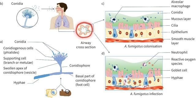
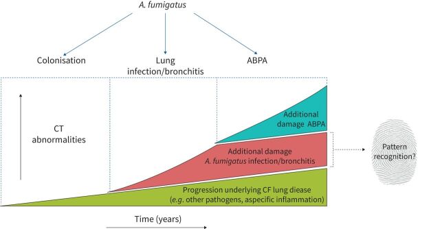
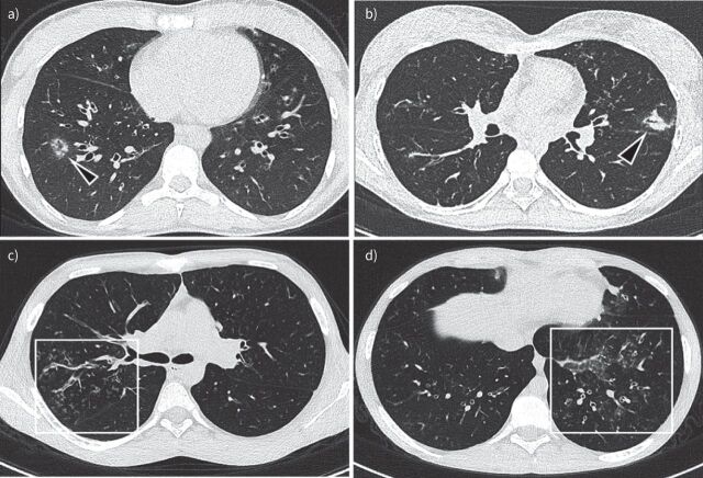
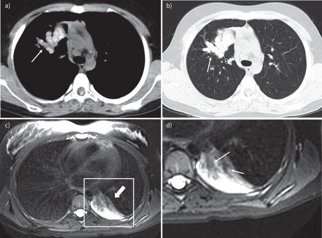
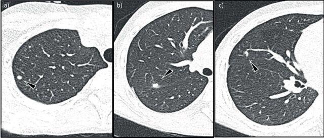
In people with cystic fibrosis (PwCF), viscous sputum and dysfunction of the mucociliary escalator leads to early and chronic infections. The prevalence of Aspergillus fumigatus in sputum is high in PwCF and the contribution of A. fumigatus to the progression of structural lung disease has been reported. However, overall, relatively little is known about the contribution of A. fumigatus to CF lung disease. More knowledge is needed to aid clinical decisions on whether to start antifungal treatment. In this review, we give an overview of A. fumigatus colonisation and infection in PwCF and the different types of pulmonary disease caused by it. Furthermore, we discuss the current evidence for structural lung damage associated with A. fumigatus in PwCF on chest computed tomography and magnetic resonance imaging. We conclude that radiological outcomes to identify disease caused by A. fumigatus can be important for clinical studies and management.


Conflict of interest statement

Conflict of interest: Q. Lv has nothing to disclose. Conflict of interest: B.B.L.J. Elders has nothing to disclose. Conflict of interest: A. Warris has nothing to disclose. Conflict of interest: D. Caudri has nothing to disclose. Conflict of interest: P. Ciet reports personal fees from Vertex Pharmaceutical, outside the submitted work. Conflict of interest: H. Tiddens reports grants and other funding from Novartis, grants from CFF and Vectura, and personal fees from Vertex, Thirona and Insmed, outside the submitted work. In addition, Erasmus MC and Telethon Kids Institute have licensed the use of PRAGMA-CF to Thirona and Resonance Health. The Sophia research BV of the Erasmus MC-Sophia Childrens hospital has received unconditional research grants from Novartis, and Vectura.

Figures

FIGURE 1
FIGURE 1
Aspergillus fumigatus from transmission to colonisation or infection. a) Microscopic morphology of Aspergillus which can grow on dead wood or starchy foods [29]. b) Inhalation of conidia into airways. Panel b) is reproduced and modified from [29] with permission. c) Colonisation of A. fumigatus in the airways in people with cystic fibrosis, where due to impaired mucociliary clearance of the thick mucus layer, the inhaled conidia are not eliminated. d) A. fumigatus infection in which germination of conidia leads to hyphal growth and tissue invasion triggering neutrophil recruitment and the release of reactive oxygen species which mediate a multitude of inflammatory pathways causing damage to the airways.
FIGURE 2
FIGURE 2
The distinction between cystic fibrosis (CF)-related and Aspergillus fumigatus-related structural lung damage in people with CF. The figure represents the difficulty of distinguishing “normal” progression of CF-related lung disease on chest computed tomography (CT) from A. fumigatus infection/bronchitis-related progression and allergic bronchopulmonary aspergillosis (ABPA)-related structural lung disease on top of this progression, and our goal to find the fingerprint for Aspergillus-related lung damage. Note: ABPA and Aspergillus bronchitis can either co-exist or occur separately.
FIGURE 3
FIGURE 3
Chest imaging of a 13 -year-old boy with cystic fibrosis. a) Magnetic resonance imaging (MRI) scan (isotropic T2-weighted CUBE, coronal reformat) and b) computed tomography (CT) scan (isotropic coronal reformat) performed 10 months after the MRI. Note cavitary lesion in the left upper lobe on MRI (arrow) with wall thickening but without a solid component. On the follow-up CT, there is a new round solid lesion within the cavity, representing an aspergilloma (arrow) not completely filling the cavity (Modod sign).
FIGURE 4
FIGURE 4
Aspergillus bronchitis/bronchiolitis example images: a) 18-year-old male non-cystic fibrosis (CF) patient, but with sickle cell disease. Multiple nodules with ground glass opacity around peripheral vessels are shown in the right lower lobe (arrowhead in a). b) Chest computed tomography (CT) image of a 17-year-old girl with CF shows Aspergillus bronchopneumonia. Note bronchocentric peri-bronchiolar consolidation on left upper lobe; c and d) Chest CT images of a 15-year-old boy with CF show Aspergillus bronchiolitis. Note filling of peripheral airways with a tree-in-bud pattern and bronchial wall thickening (box in c). In d there is also an area of ground glass (box in d).
FIGURE 5
FIGURE 5
Allergic bronchopulmonary aspergillosis example images. a) and b) Chest computed tomography (CT) images of a 12-year-old boy with cystic fibrosis (CF). a) 2.5 mm axial CT soft tissue window and b) 1 mm axial CT lung window at the same level. Note in a), central bronchiectasis filled with high density material (arrow). In b), note mucus plugs with a typical cylindrical shape originating from the hilar region and extending towards the periphery (arrow) showing typical higher density than paraspinal muscles. c) and d) Lung magnetic resonance imaging scans (T2-weighted axial PROPELLER, in plane resolution 1×1 mm, slice thickness 5 mm) of a 15-year-old girl with CF. c) Atelectasis in the left lower lobe (thick arrow). d) Hypo-intense material filling the central airways (arrows).
FIGURE 6
FIGURE 6
Chronic aspergillosis with skin and lung abnormality in a 15-year-old boy with cardiomyopathy. Note sub-pleural (arrowhead in a) and peri-fissural (arrowheads in b and c) nodules. From their location, these nodules are most likely intrapulmonary lymph nodes, which disappeared after 4 months of treatment with antifungal medication (voriconazole).

References

    1. O'Sullivan BP, Freedman SD. Cystic fibrosis. Lancet 2009; 373: 1891–1904. doi:10.1016/S0140-6736(09)60327-5 - DOI - PubMed
    1. McMahon MA, Chotirmall SH, McCullagh B, et al. . Radiological abnormalities associated with Aspergillus colonization in a cystic fibrosis population. Eur J Radiol 2012; 81: e197–e202. doi:10.1016/j.ejrad.2011.01.114 - DOI - PubMed
    1. Ratjen F, Doring G. Cystic fibrosis. Lancet 2003; 361: 681–689. doi:10.1016/S0140-6736(03)12567-6 - DOI - PubMed
    1. Foster K, Alton H. Chronic lung infection in children. Paediatr Respir Rev 2003; 4: 225–229. doi:10.1016/S1526-0542(03)00054-X - DOI - PubMed
    1. King J, Brunel SF, Warris A. Aspergillus infections in cystic fibrosis. J Infect 2016; 72: Suppl., S50–S55. doi:10.1016/j.jinf.2016.04.022 - DOI - PubMed

Substances