. 2021 Nov 17;11(1):22434.
doi: 10.1038/s41598-021-01790-6.

Single-cell RNA sequencing reveals differential cell cycle activity in key cell populations during nephrogenesis

Affiliations

Single-cell RNA sequencing reveals differential cell cycle activity in key cell populations during nephrogenesis

Abha S Bais et al. Sci Rep. .

Abstract

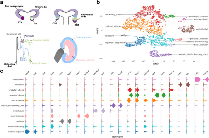
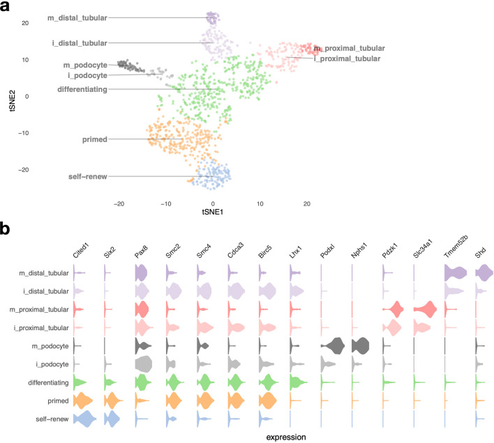
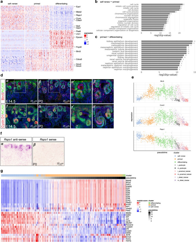
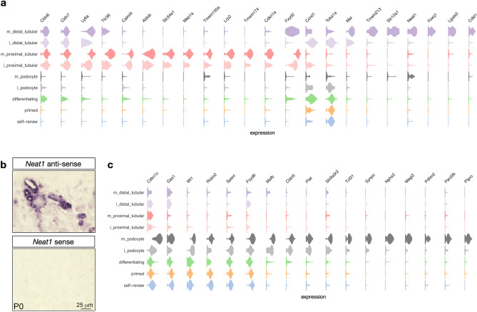
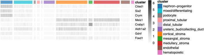
The kidney is a complex organ composed of more than 30 terminally differentiated cell types that all are required to perform its numerous homeostatic functions. Defects in kidney development are a significant cause of chronic kidney disease in children, which can lead to kidney failure that can only be treated by transplant or dialysis. A better understanding of molecular mechanisms that drive kidney development is important for designing strategies to enhance renal repair and regeneration. In this study, we profiled gene expression in the developing mouse kidney at embryonic day 14.5 at single-cell resolution. Consistent with previous studies, clusters with distinct transcriptional signatures clearly identify major compartments and cell types of the developing kidney. Cell cycle activity distinguishes between the "primed" and "self-renewing" sub-populations of nephron progenitors, with increased expression of the cell cycle-related genes Birc5, Cdca3, Smc2 and Smc4 in "primed" nephron progenitors. In addition, augmented expression of cell cycle related genes Birc5, Cks2, Ccnb1, Ccnd1 and Tuba1a/b was detected in immature distal tubules, suggesting cell cycle regulation may be required for early events of nephron patterning and tubular fusion between the distal nephron and collecting duct epithelia.


Conflict of interest statement

The authors declare no competing interests. Our study with observational experimental design was carried out in compliance with the ARRIVE guidelines.

Figures

Figure 1
Figure 1
Developing embryonic day 14.5 mouse kidney cell types. (a) Schematic illustration of nephron induction and patterning. In response to signals from the ureteric bud, the metanephric mesenchyme condenses and forms a cap of nephron progenitors (= cap mesenchyme) around the ureteric bud tips. Next, a sub-population of nephron progenitors undergoes a mesenchymal to epithelial transition to form pre-tubular aggregates (PTA), which develop sequentially into renal vesicles (RV), comma-shaped body (CSB) and S-shaped body (SSB). Endothelial cells are attracted into the cleft of the SSB. Color-coded map indicates the cell fate relationship of progenitor regions in SSB structure (upper right) and adult nephron structure (lower left). Schematic of a lateral view of the metanephric kidney depicting the cortical and medullary stroma (lower right). (b) tSNE plot showing the eleven cell clusters in the embryonic mouse kidney, with cell clusters corresponding to major components indicated by color. (c) Violin plots of gene expression for known lineage-associated genes (columns), stratified by cluster (rows). Our data clearly identifies cells from the major structural components of the developing kidney.
Figure 2
Figure 2
Cell types of the nephron progenitor lineage. (a) shows a tSNE plot of NP-derived cells, with clusters corresponding to cell types indicated by colors. The prefix “i_” indicates immature cells, while “m_” indicates mature cells. (b) shows violin plots of gene expression for known lineage-associated genes (columns), stratified by cluster (rows). We observe two types of NP cells (“self-renewing” and “primed”) and clear separation of distal and proximal tubular cells and podocytes in our data.
Figure 3
Figure 3
Transcriptional signatures of self-renewing, primed and differentiating nephron progenitor cells. (a) shows differentially expressed genes on a heatmap of 100 random cells for each of the “self-renew”, “primed” and differentiating clusters, with key genes annotated on the right. (b) and (c) show the 20 most-enriched Gene Ontology terms for genes differentially expressed between self-renewing and primed NP cells, and between primed NP cells and differentiating cells, respectively. (d) Immunofluorescence on kidney sections from embryonic day 14.5 (E14.5) and postnatal day 0 (P0) mice using anti-BIRC5 (α - β′) and anti-Cyclin D1 (γ - δ’) antibodies (red). Nephron progenitors and their early epithelial derivatives were detected using an antibody against anti-Neural cell adhesion molecule (NCAM; green). Nuclei were counterstained with DAPI (blue). Scale bar, 25 μm. The sub-panels α′, β′, γ′ and δ′ are close-ups of the areas indicated by the white boxes. (e) Expression of Birc5, Ccnd1 and Rspo1 across pseudotime; colors indicate cell clusters. (f) In situ hybridization on cryosections of P0 kidneys confirms the expression of Rspo1 in nephron progenitors and their early epithelial derivatives (α). No signal was detected with sense probe hybridization (β). Images are representative of three independent experiments. Scale bar 25 μm. (g) Inferred regulatory module activity based on SCENIC across pseudotime for predominantly self-renewing and primed nephron progenitor cells.
Figure 4
Figure 4
Transcriptional signatures of podocytes and tubular cells. (a) Violin plot of genes expressed in proximal and distal tubular cells (rows are clusters and columns denote genes). (b) In situ hybridization of P0 kidneys confirms expression of Neat1 in distal tubules. No signal was detected with sense probe hybridization. (c) Same as (a), but for podocytes.
Figure 5
Figure 5
Expression of lineage-marker genes in unexpected cell types. Heatmap of gene expression (gray scale) of known lineage marker genes (rows) across cells (columns), ordered by cell clusters (color index). We observe the expression of cap mesenchyme markers (Cited1, Six2, Crym, Gdnf) in stromal cells and vice versa, consistent with previous reports.

Similar articles

Cited by

References

    1. Capone VP, Morello W, Taroni F, Montini G. Genetics of congenital anomalies of the kidney and urinary tract: the current state of play. Int. J. Mol. Sci. 2017;18:796. doi: 10.3390/ijms18040796. - DOI - PMC - PubMed
    1. Yosypiv IV. Congenital anomalies of the kidney and urinary tract: a genetic disorder? Int. J. Nephrol. 2012;2012:909083. doi: 10.1155/2012/909083. - DOI - PMC - PubMed
    1. Keller G, Zimmer G, Mall G, Ritz E, Amann K. Nephron number in patients with primary hypertension. N Engl. J. Med. 2003;348:101–108. doi: 10.1056/NEJMoa020549. - DOI - PubMed
    1. Hoy WE, Hughson MD, Singh GR, Douglas-Denton R, Bertram JF. Reduced nephron number and glomerulomegaly in Australian Aborigines: a group at high risk for renal disease and hypertension. Kidney Int. 2006;70:104–110. doi: 10.1038/sj.ki.5000397. - DOI - PubMed
    1. Humphreys BD. Mechanisms of Renal Fibrosis. Annu Rev Physiol. 2018;80:309–326. doi: 10.1146/annurev-physiol-022516-034227. - DOI - PubMed

Publication types