Case Reports
. 2021 Dec;600(7887):133-137.
doi: 10.1038/s41586-021-04111-z. Epub 2021 Nov 17.

Independent infections of porcine deltacoronavirus among Haitian children

Affiliations
Case Reports

Independent infections of porcine deltacoronavirus among Haitian children

John A Lednicky et al. Nature. 2021 Dec.

Abstract

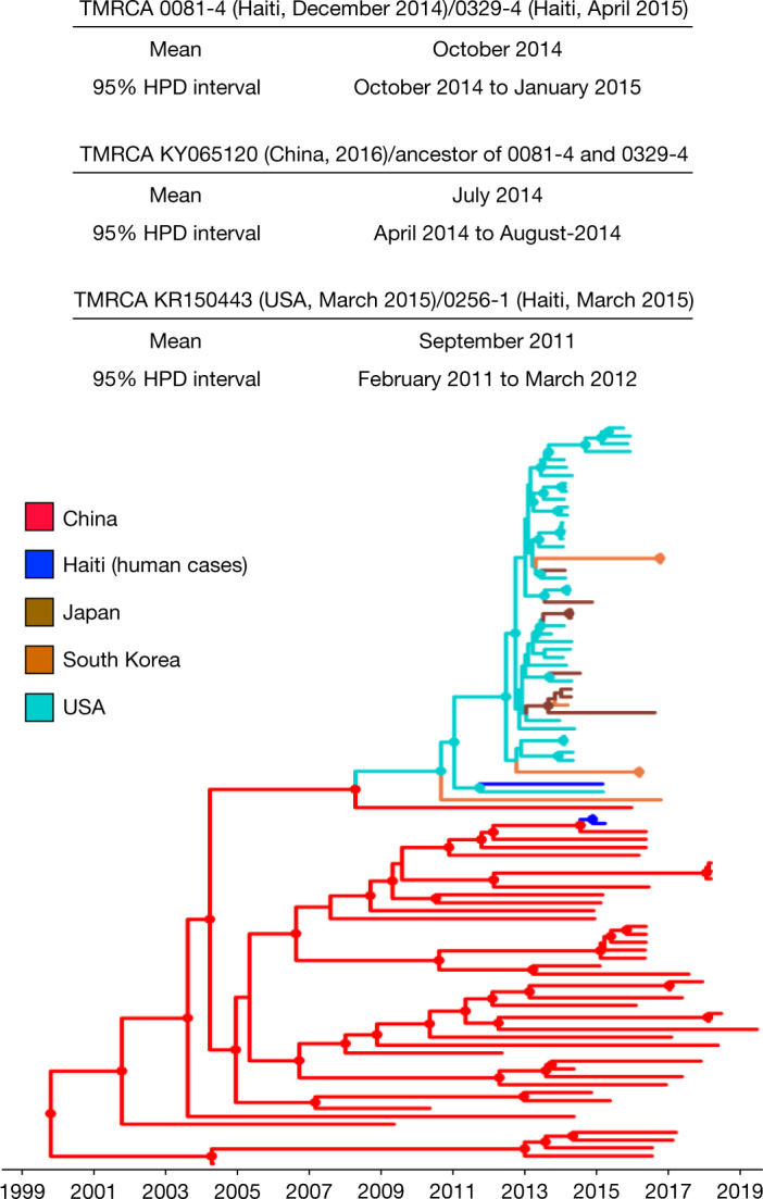
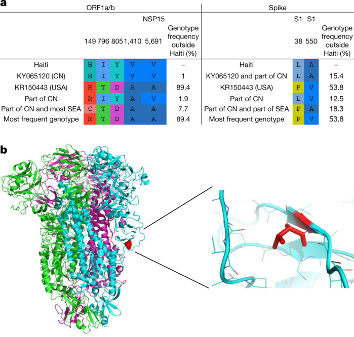
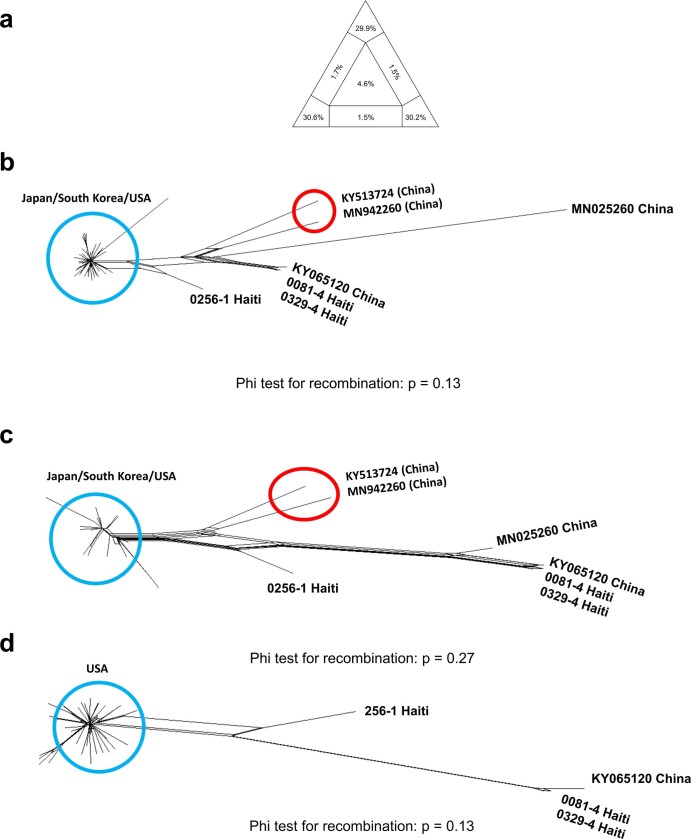
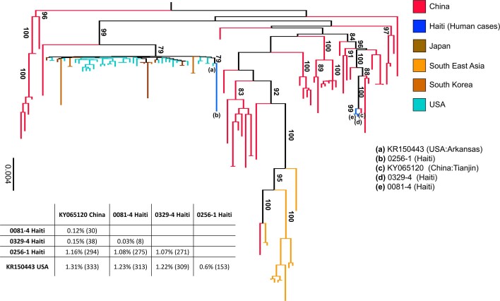
Coronaviruses have caused three major epidemics since 2003, including the ongoing SARS-CoV-2 pandemic. In each case, the emergence of coronavirus in our species has been associated with zoonotic transmissions from animal reservoirs1,2, underscoring how prone such pathogens are to spill over and adapt to new species. Among the four recognized genera of the family Coronaviridae, human infections reported so far have been limited to alphacoronaviruses and betacoronaviruses3-5. Here we identify porcine deltacoronavirus strains in plasma samples of three Haitian children with acute undifferentiated febrile illness. Genomic and evolutionary analyses reveal that human infections were the result of at least two independent zoonoses of distinct viral lineages that acquired the same mutational signature in the genes encoding Nsp15 and the spike glycoprotein. In particular, structural analysis predicts that one of the changes in the spike S1 subunit, which contains the receptor-binding domain, may affect the flexibility of the protein and its binding to the host cell receptor. Our findings highlight the potential for evolutionary change and adaptation leading to human infections by coronaviruses outside of the previously recognized human-associated coronavirus groups, particularly in settings where there may be close human-animal contact.


Conflict of interest statement

The authors declare no competing interests.

Figures

Fig. 1
Fig. 1. Non-specific CPEs formed by plasma from patient 0081-4 in Vero E6 cells.
a, Mock-infected Vero E6 cells, 11 days post-inoculation with phosphate-buffered saline. b, Vero E6 cells 11 days post-inoculation with plasma from patient 0081-4. Original magnification, ×200.
Fig. 2
Fig. 2. Bayesian maximum clade credibility tree of PDCoV strains.
The circles at the internal nodes indicate high posterior probability support of more than 0.9. The branch lengths were scaled in time by using a strict molecular clock. The information above the tree shows the inferred TMRCA between strains discovered in Haiti and their closest phylogenetic relative, with 95% HPD intervals.
Fig. 3
Fig. 3. Analysis of conserved amino acids in human PDCoV strains.
Residues are numbered based on the ORF1a/b absolute amino acid position in the reference sequence JQ065043 (pig/China). a, Amino acid signature pattern analysis on ORF1a/b. CN, China; SEA, southeast Asia. b, The trimer structure of the spike glycoprotein. Different colours are assigned to each monomer: red indicates residue 550, where Haitian sequences have a valine to alanine mutation.
Extended Data Fig. 1
Extended Data Fig. 1. Negative stained coronavirus particle viewed by transmission electron microscopy.
Typical coronavirus spikes rim the spherical virus particle.
Extended Data Fig. 2
Extended Data Fig. 2. Assessment of phylogenetic signal and Neighbor net plot of PDCoV fragments.
a, Likelihood mapping of the final 109 sequences alignment (see Methods) showing extremely low phylogenetic noise (4.6% in the center of the triangle), as required for reliable phylogeny inference. b, Neighbor net inferred from pair-wise p-distances of 47 genomes for the major genome fragment identified by RDP4 (see Methods). Recombination signal was assessed by the PHI test. No significant evidence of recombination was found in either fragment (PHI test p > 0.05). c, Neighbor net and PHI test for the minor genome fragment identified by RDP4 (see Methods). The only change in topology, in the split decomposition networks shown in panels a and b, concerned two Chinese sequences (highlighted by the red circle) displaying mixed ancestry possibly due to homoplasy. These sequences were also removed before performing any further phylogenetic analyses. d, Neighbor net and PHI test for the full genome alignment only including Haitian Hu-PDCoV strains and closest Chinese and American strains (after removal recombinant strains detected by RDP4).
Extended Data Fig. 3
Extended Data Fig. 3. Maximum likelihood tree of PDCoV strains.
The tree was inferred from 109 PDCoV full genome sequences, including four sparrow CoV genomes for outgroup rooting (accession numbers MG812375, MG812376, MG812377, and MG812378, from Chen et al.) using the best fitting nucleotide substitution model (see Methods). For display purposes, outgroup sequences were removed from the figure. Vertical branches are scaled in number of nucleotide substitutions per site according to the bar on the left of the tree. Bootstrap values > 75% (1000 replicates) are indicated along supported branches. The table at the bottom of the tree shows nucleotide % dissimilarity (and total number of nucleotide differences in parenthesis) between Haitian strains and their closest non-Haitian relatives, which are labelled in tree by progressive letters according to the legend on the right.
Extended Data Fig. 4
Extended Data Fig. 4. Analysis of the temporal signal with TempEst.
a, b, Root-to-tip distance (y-axis) vs. sampling time linear regression in the ML likelihood phylogeny inferred from: all PDCoV sequences in the final data set (n = 109 sequences) including sparrow outgroup sequences (a); PDCoV sequences without outgroups (n = 105 sequences), with sequences in the red circle belonging to the South East Asia clade (see Fig. 1) showing a clear departure from the strict molecular clock model (b); and PDCoV sequences after removal of the South East Asia clade (n = 94 sequences), showing greatly improved clock signal (R2 = 0.68) (c).
Extended Data Fig. 5
Extended Data Fig. 5. Bayesian maximum clade credibility (MCC) tree of PDCoV strains.
The MCC tree was evolutionary rate. Circles at internal nodes indicate high posterior probability (PP) support >0.9. The table on inferred from a subset of 94 full genome strains that displayed sufficient temporal signal for molecular clock calibration. Branch lengths were scaled in time, according to the bar at the bottom, by using a strict molecular clock and sampling dates to estimate PDCoV the left shows the inferred time of the most recent common ancestor (TMRCA) between Haitian strains and their closest phylogenetic relative, with 95% high posterior density intervals (95%HPD).

Update of

References

    1. Su S, et al. Epidemiology, genetic recombination, and pathogenesis of coronaviruses. Trends Microbiol. 2016;24:490–502. - PMC - PubMed
    1. Wu F, et al. A new coronavirus associated with human respiratory disease in China. Nature. 2020;579:265–269. - PMC - PubMed
    1. Ye ZW, et al. Zoonotic origins of human coronaviruses. Int. J. Biol. Sci. 2020;16:1686–1697. - PMC - PubMed
    1. Fehr AR, Perlman S. Coronaviruses: an overview of their replication and pathogenesis. Methods Mol. Biol. 2015;1282:1–23. - PMC - PubMed
    1. Graham RL, Baric RS. Recombination, reservoirs, and the modular spike: mechanisms of coronavirus cross-species transmission. J. Virol. 2010;84:3134–3146. - PMC - PubMed

Publication types

MeSH terms