. 2022 Jul;43(7):1780-1792.
doi: 10.1038/s41401-021-00797-z. Epub 2021 Nov 17.

Ainsliadimer C, a disesquiterpenoid isolated from Ainsliaea macrocephala, ameliorates inflammatory responses in adipose tissue via Sirtuin 1-NLRP3 inflammasome axis

Affiliations

Ainsliadimer C, a disesquiterpenoid isolated from Ainsliaea macrocephala, ameliorates inflammatory responses in adipose tissue via Sirtuin 1-NLRP3 inflammasome axis

Cheng Chen et al. Acta Pharmacol Sin. 2022 Jul.

Abstract

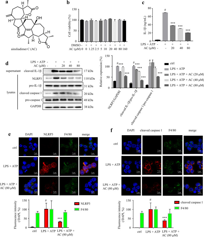
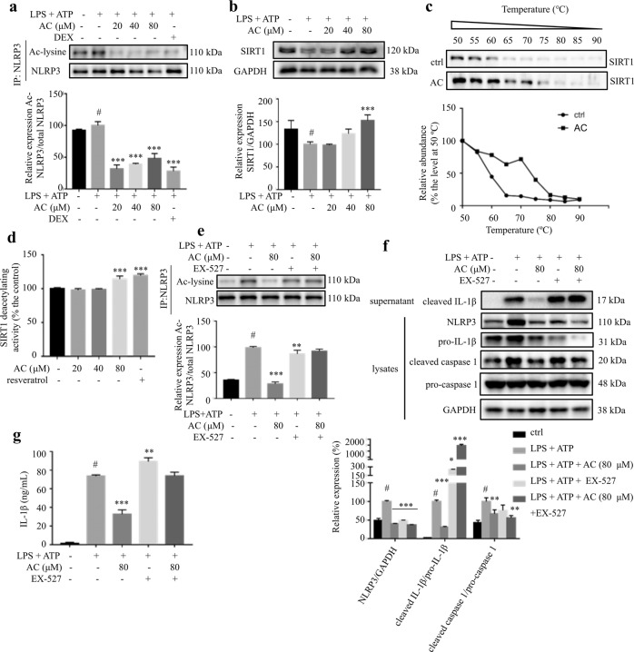
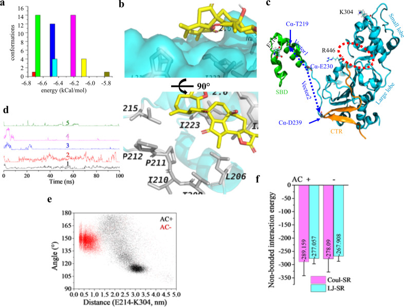
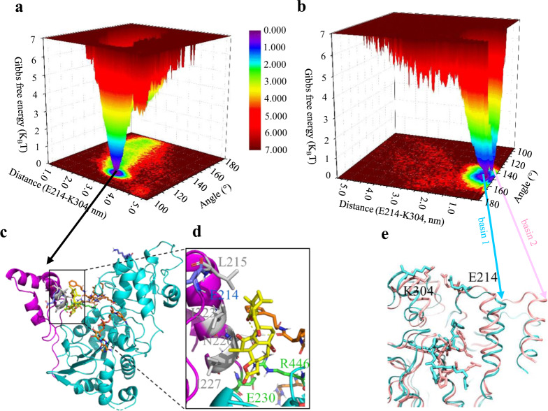
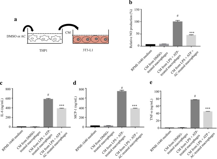
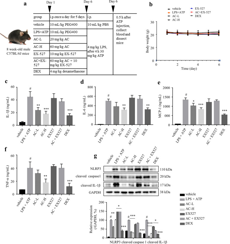
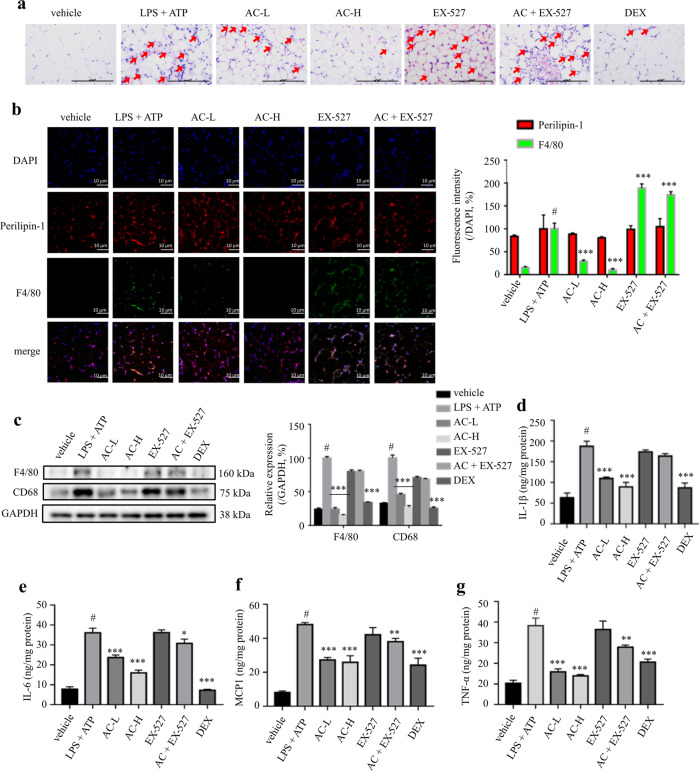
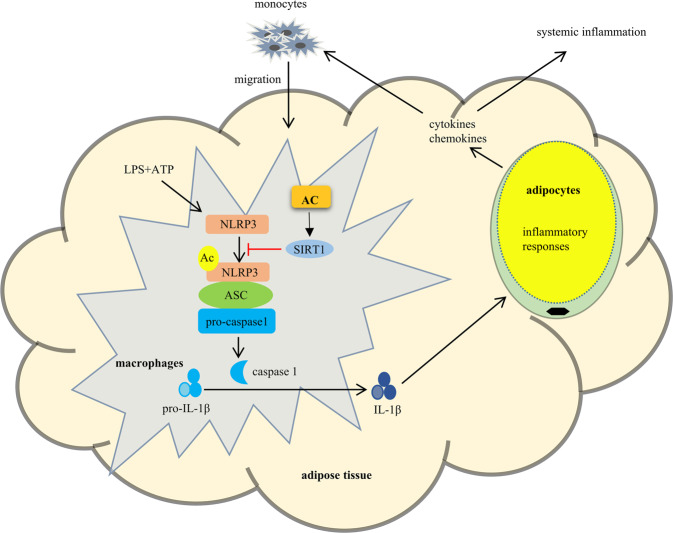
Interleukin-1β (IL-1β), a key pro-inflammatory cytokine, is majorly produced by macrophages through NOD-, LRR-, and pyrin domain-containing protein 3 (NLRP3) inflammasome, which has been identified as the culprit to deteriorate the inflammatory crosstalk between macrophages and adipocytes. Ainsliadimer C (AC) is a disesquiterpenoid isolated from Ainsliaea macrocephala. In the current study, we investigated the effects of AC on adipose tissue inflammation in co-culture of macrophages and adipocytes in vitro as well as in LPS-treated mice in vivo. We showed that AC (20-80 µM) dose-dependently inhibited the secretion of IL-1β from LPS plus ATP-stimulated THP-1 macrophages by inhibiting the activation of NLRP3 inflammasome. Furthermore, we found that AC treatment activated NAD+-dependent deacetylase Sirtuin 1 (SIRT1), resulting in reduced acetylation level of NLRP3. Molecular modeling analysis revealed that binding of AC to sirtuin-activating compound-binding domain increased the affinity of the substrate to the catalytic domain of SIRT1. Moreover, AC (80 µM) significantly attenuated macrophage-conditioned medium-induced inflammatory responses in 3T3-L1 adipocytes. In LPS-induced acute inflammatory mice, administration of AC (20, 60 mg·kg-1·d-1, ip) for 5 days significantly suppressed the pro-inflammatory cytokine levels in serum and epididymal white adipose tissue (eWAT), attenuated macrophage infiltration into eWAT, and mitigated adipose tissue inflammation. The beneficial effects of AC were blocked by co-administration of a selective SIRT1 inhibitor EX-527 (10 mg·kg-1·d-1). Taken together, AC suppresses NLRP3-mediated IL-1β secretion through activating SIRT1, leading to attenuated inflammation in macrophages and adipose tissue, which might be a candidate to treat obesity-associated metabolic diseases.

Keywords: Ainsliadimer C; EX-527; NLRP3 inflammasome; Sirtuin 1; adipose tissue inflammation; dexamethasone; interleukin-1β; macrophages; metabolic disorders; obesity.


Conflict of interest statement

The authors declare no competing interests.

Figures

Fig. 1
Fig. 1. AC suppressed LPS plus ATP-stimulated IL-1β secretion in THP-1 cells.
a Chemical structure of AC. b Cytotoxicity of AC on THP-1 cells assessed by MTT assay. THP-1 cells were treated with different concentrations of AC (from 1.25 to 160 μM) for 24 h (n = 6). c The levels of IL-1β in the culture medium from THP-1 cells (n = 3). THP-1 cells were cultured in the presence or absence of AC for 12 h, and stimulated with LPS for 4 h and then ATP for 1 h. d The expression of NLRP3, pro-caspase 1, cleaved caspase 1, pro-IL-1β in the lysates, and cleaved IL-1β in the supernatant of THP-1 cells detected by Western blotting. GAPDH was chosen as an internal loading control (n = 5). Immunofluorescence staining of NLRP3 (e) and cleaved caspase 1 (f) (n = 3). Scale bar = 10 μm. Data are expressed as means ± SEM. #P < 0.05 versus DMSO, ***P < 0.001 versus LPS + ATP.
Fig. 2
Fig. 2. AC inhibited the activation of NLRP3 inflammasome through activating SIRT1.
THP-1 cells were treated with various concentrations of AC for 12 h, and then stimulated with LPS for 4 h and ATP for 1 h. a The acetylated and total NLRP3 protein levels (n = 3). b The expression level of SIRT1 detected by Western blotting (n = 3). GAPDH was chosen as an internal loading control. c CETSA performed on THP-1 cells after the treatment with or without AC (80 μM) for 12 h (n = 3). d The effect of AC or resveratrol on the deacetylating activity of SIRT1 (n = 6). The deacetylating activity was determined with a SIRT1 fluorometric assay kit. e The acetylated and total NLRP3 protein levels (n = 3). f The expression of NLRP3, pro-caspase 1, cleaved caspase 1, and pro-IL-1β in the lysates, and cleaved IL-1β in the supernatant of THP-1 cells detected by Western blotting (n = 3). GAPDH was chosen as an internal loading control. g The levels of IL-1β in the culture medium from THP-1 cells (n = 6). Data are expressed as means ± SEM. #P < 0.05 versus DMSO, **P < 0.01, ***P < 0.001 versus LPS + ATP.
Fig. 3
Fig. 3. AC is a potential activator of SIRT1.
a Docking results of AC to the reported allosteric activation site of SIRT1 (PDB ID: 4ZZJ). Cluster analysis of the acquired conformations from AutoDock 4.2 using a tolerance of 2.0 Å. b Interactions between AC and residues on SIRT1 of the most possible binding conformation. Residues and AC are shown in white gray and yellow sticks. Hydrogen bond is indicated in yellow dashed line. c SIRT1 structure showing key domains, amino acid pairs, and the two vectors monitored. Protein structure was shown in NewCartoon, E214 and K304 were shown in CPK, and R446 was displayed as Licorice; the red dashed circle indicates the catalytic cleft; α-carbon atoms were shown in VDW; vector defined from the Cα of E230 to that of T219 was termed as vector1 and to that of D239 was termed as vector 2. d Changes of the distance as a function of time between the carboxyl oxygen atoms of E230 and the nitrogen atoms on the guanidyl group of R446 from five independent dynamic simulations in the holo structure. e Scatter plots showing the conformation distribution in the presence/absence of AC using the angle from the two vectors and the distance between E214 and K304. f Non-bonded interaction energy between the substrate and the catalytic domain calculated from the stable trajectories of the holo structure and that in state 2 of the apo structure. Coul-SR short-range Coulomb interaction energy, LJ-SR short-range Lennard–Jones potential.
Fig. 4
Fig. 4. Free energy landscape and representative conformations in the presence/absence of AC.
a, b Free energy landscape of the holo structure and apo structure. Free energy surfaces are colored as gradient, where the units are expressed as KBT. KB is the Boltzmann constant and T isthe temperature. ce Representative conformation in the energy basins of the holo structure and the apo structure. The SBD and catalytic domain were colored as magenta and cyan, respectively; residues, AC (yellow), NAD, and the substrate were shown in sticks; hydrogen bond was indicated as yellow dashed line.
Fig. 5
Fig. 5. AC attenuated macrophage-CM-induced inflammatory responses in adipocytes.
a Schematic diagram of the experimental procedure. THP-1 cells were treated AC (80 μM) or DMSO for 12 h, and subsequently stimulated with LPS for 4 h and ATP (1 mM) for 1 h. After 24 h of culturing in fresh medium, the supernatants were collected as macrophage CM, which was used to incubate the fully differentiated 3T3-L1 for 24 h. The production of NO (b) and the levels of IL-6 (c), MCP-1 (d), and TNF-α (e) in 3T3-L1 adipocytes were determined. Data are expressed as means ± SEM. n = 6. #P < 0.05 versus DMSO, ***P < 0.001 versus LPS + ATP.
Fig. 6
Fig. 6. AC treatment alleviated LPS plus ATP-mediated inflammatory responses in mice.
a The inducement of in vivo acute inflammation model using LPS plus ATP. DEX: 4 mg/kg DEX; AC-L: 20 mg/kg AC; AC-H: 60 mg/kg AC; EX-527: 10 mg/kg EX-527; AC + EX-527: 60 mg/kg AC and 10 mg/kg EX-527. n = 3‒5. b Changes of body weight. The serum levels of IL-1β (c), IL-6 (d), MCP-1 (e), and TNF-α (f). g NLRP3, cleaved IL-1β, and cleaved caspase 1 levels in peritoneal macrophages were analyzed by Western blotting. GAPDH was chosen as an internal loading control. Data are expressed as means ± SEM. #P < 0.05 LPS + ATP vs. vehicle; *P < 0.05, **P < 0.01, ***P < 0.001 AC-L, AC-H or DEX vs. LPS + ATP.
Fig. 7
Fig. 7. AC treatment attenuated adipose tissue inflammation.
a H&E staining of eWAT sections. Red arrows indicate the infiltrated macrophages in eWAT. Scale bar = 100 μm. b Immunofluorescence staining of perilipin-1 (red, marker of adipocytes) and F4/80 (green, marker of macrophages) in eWAT. Scale bar = 10 μm. c Expression levels of F4/80 and CD68 determined by western blotting. GAPDH was chosen as an internal loading control. The levels of IL-1β (d), IL-6 (e), MCP-1 (f), and TNF-α (g) in eWAT determined by ELISA. DEX: 4 mg/kg DEX; AC-L: 20 mg/kg AC; AC-H: 60 mg/kg AC; EX-527: 10 mg/kg EX-527; AC + EX-527: 60 mg/kg AC and 10 mg/kg EX-527. Data are expressed as means ± SEM (n = 3‒5). #P < 0.05 LPS + ATP vs. vehicle; *P < 0.05, **P < 0.01, ***P < 0.001 AC-L, AC-H or DEX vs. LPS + ATP.
Fig. 8
Fig. 8. Schematic model of AC in attenuating adipose tissue inflammation.
Therapeutically, AC activates SIRT1 to suppress the activation of NLRP3 inflammasome, which in turn alleviates macrophages and adipose tissue inflammation by reducing the secretion of IL-1β.

Similar articles

Cited by

References

    1. Ellulu MS, Patimah I, Khaza′ai H, Rahmat A, Abed Y. Obesity and inflammation: the linking mechanism and the complications. Arch Med Sci. 2017;4:851–63. doi: 10.5114/aoms.2016.58928. - DOI - PMC - PubMed
    1. Lee CH. Young eosinophils rejuvenate ageing adipose tissues. Nat Metab. 2020;2:655–6. doi: 10.1038/s42255-020-0230-9. - DOI - PubMed
    1. Swanson KV, Deng M, Ting JP. The NLRP3 inflammasome: molecular activation and regulation to therapeutics. Nat Rev Immunol. 2019;19:477–89. doi: 10.1038/s41577-019-0165-0. - DOI - PMC - PubMed
    1. Li D, Liu Q, Sun W, Chen X, Wang Y, Sun Y, et al. 1,3,6,7-Tetrahydroxy-8-prenylxanthone ameliorates inflammatory responses resulting from the paracrine interaction of adipocytes and macrophages. Br J Pharmacol. 2018;175:1590–606. doi: 10.1111/bph.14162. - DOI - PMC - PubMed
    1. Zhang T, Fang Z, Linghu KG, Liu J, Gan L, Lin L. Small molecule-driven SIRT3-autophagy-mediated NLRP3 inflammasome inhibition ameliorates inflammatory crosstalk between macrophages and adipocytes. Br J Pharmacol. 2020;177:4645–65. doi: 10.1111/bph.15215. - DOI - PMC - PubMed