

Solitary Osteochondromas of the Metatarsal and Cuneiform, in an Adolescent
- PMID: 34790613
- PMCID: PMC8576773
- DOI: 10.13107/jocr.2021.v11.i07.2332
Solitary Osteochondromas of the Metatarsal and Cuneiform, in an Adolescent
Abstract
Introduction: Solitary osteochondromas are extremely rare in the bones of the foot. In the growing skeleton, few cases affecting the metatarsals and the talus have been reported. At present, there have been no reports of osteochondromas affecting the cuneiforms.
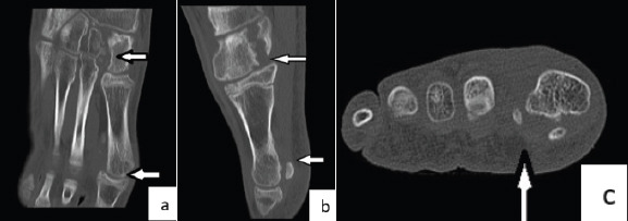
Case report: We report the case of a 13-year-old male patient. He presented with marked prominences in the plantar surface of his left foot and pain while participating in sporting activities. Radiological examination with X-rays, computed tomography (CT) scan, and magnetic resonance imaging revealed two solitary osteochondromas growing from the medial cuneiform and the head of the 1st metatarsal. The patient was treated surgically by excision of the osteochondromas. Histological examination confirmed the diagnosis of osteochondromas. He had an uneventful recovery and returned to his sporting activities.
Conclusion: Solitary osteochondroma can present in the cuneiform and metatarsal of a growing adolescent. CT scan is useful for the accurate diagnosis and surgical removal of the tumor.
Keywords: Osteochondroma; child; cuneiform; foot; metatarsal.
Copyright: © Indian Orthopaedic Research Group.
Conflict of interest statement
Conflict of Interest: Nil
Figures


References
-
- Fuselier CO, Binning T, Kushner D, Kirchwehm WW, Rice JR, Hetherington V, et al. Solitary osteochondroma of the foot:An in-depth study with case reports. J Foot Surg. 1984;23:3–24. - PubMed
-
- Turati M, Bigoni M, Omeljaniuk RJ, Griffet J, Zatti G, Courvoisier A. Pediatric navicular dorsal osteochondroma:A rare case of navicular-cuneiform impingement. J Pediatr Orthop B. 2019;28:602–6. - PubMed
-
- Blitz N, Lopez K. Giant solitary osteochondroma of the inferior medial calcaneal tubercle:A case report and review of the literature. J Foot Ankle Surg. 2008;47:206–12. - PubMed
-
- Patil SD, Patil VD, Khan A, Khanore C. Correction of a forefoot deformity caused by a large, solitary metatarsal osteochondroma in an adolescent:A case report. J Foot Ankle Surg. 2016;55:427–33. - PubMed
Publication types
LinkOut - more resources
Full Text Sources