Review
. 2021 Nov 1:9:723532.
doi: 10.3389/fped.2021.723532. eCollection 2021.

Autoimmune Polyendocrinopathy-Candidiasis-Ectodermal Dystrophy

Affiliations
Review

Autoimmune Polyendocrinopathy-Candidiasis-Ectodermal Dystrophy

Elise M N Ferré et al. Front Pediatr. .

Abstract

Autoimmune polyendocrinopathy-candidiasis-ectodermal dystrophy (APECED), also known as autoimmune polyglandular syndrome type-1 (APS-1), is a rare monogenic autoimmune disease caused by loss-of-function mutations in the autoimmune regulator (AIRE) gene. AIRE deficiency impairs immune tolerance in the thymus and results in the peripheral escape of self-reactive T lymphocytes and the generation of several cytokine- and tissue antigen-targeted autoantibodies. APECED features a classic triad of characteristic clinical manifestations consisting of chronic mucocutaneous candidiasis (CMC), hypoparathyroidism, and primary adrenal insufficiency (Addison's disease). In addition, APECED patients develop several non-endocrine autoimmune manifestations with variable frequencies, whose recognition by pediatricians should facilitate an earlier diagnosis and allow for the prompt implementation of targeted screening, preventive, and therapeutic strategies. This review summarizes our current understanding of the genetic, immunological, clinical, diagnostic, and treatment features of APECED.

Keywords: AIRE; APECED syndrome; APS-1; IFN-γ; IL-17; autoantibodies; self-reactive T cells; type-I interferons.


Conflict of interest statement

The authors declare that the research was conducted in the absence of any commercial or financial relationships that could be construed as a potential conflict of interest.

Figures
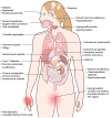
Figure 1
Figure 1
Spectrum of clinical manifestations in APECED patients. Depiction of organ-specific autoimmune manifestations observed with variable frequencies in patients with APECED. Derived from Constantine and Lionakis (13) with permission from Wiley.
Figure 2
Figure 2
Representative histological features of common non-endocrine manifestations in APECED patients. (A) A liver biopsy displaying severe chronic hepatitis with expansion of portal areas by inflammation and fibrosis and extensive interface hepatitis with numerous plasma cells infiltrating into the hepatic parenchyma (H&E; scale bar: 50 μm; original magnification, ×200). (B) A minor salivary gland biopsy depicting lymphocytic and plasma cell infiltration in and around the ducts of the gland (H&E; scale bar: 50 μm; original magnification, ×200). (C) Skin biopsy of a patient with APECED rash demonstrates perivascular and periadnexal inflammation in the superficial and deep dermis with pallor of the papillary dermis (H&E; scale bar: 2 mm; original magnification, ×40) (D) An open lung biopsy revealing chronic bronchiolitis with lymphocytic infiltration within and around the bronchiolar mucosa and lymphoid aggregates in the interstitium nearby (H&E; scale bar: 50 μm; original magnification, ×200). (E) A stomach biopsy exhibiting chronic antral inflammation with lymphoplasmacytic infiltrates in the lamina propria and occasionally on glands (H&E; scale bar: 50 μm; original magnification, ×200). (F) A jejunal biopsy depicting mild villus blunting and focal acute inflammation (H&E; scale bar: 50 μm; original magnification, ×200). Images in panels A-E are derived from Ferré et al., (7).
Figure 3
Figure 3
Clinical images depicting ophthalmologic, mucocutaneous and dermatologic manifestations in APECED patients. (A) Extensive keratopathy secondary to chronic keratoconjunctivitis. (B) Depiction of Candida chelitis and thrush affecting the tongue. (C) Endoscopic visualization of the esophagus demonstrating Candida esophagitis. (D) Nail dystrophy. (E) Alopecia areata. (F) Vitiligo affecting the knees. (G) APECED rash in an infant with APECED. Image 3G is derived from Ferré et al., (7).
Figure 4
Figure 4
Diagnostic algorithm in patients with suspected APECED. The presence of any 2 manifestations amongst the combined classic and adjunct triads should raise suspicion for APECED and should prompt further work-up with AIRE sequencing and measurement of IFNω autoantibodies.

References

    1. Finnish-German AC. An autoimmune disease, APECED, caused by mutations in a novel gene featuring two PHD-type zinc-finger domains. Nat Genet. (1997) 17:399–403. 10.1038/ng1297-399 - DOI - PubMed
    1. Nagamine K, Peterson P, Scott HS, Kudoh J, Minoshima S, Heino M, et al. . Positional cloning of the APECED gene. Nat Genet. (1997) 17:393–8. 10.1038/ng1297-393 - DOI - PubMed
    1. Anderson MS, Venanzi ES, Klein L, Chen Z, Berzins SP, Turley SJ, et al. . Projection of an immunological self shadow within the thymus by the aire protein. Science. (2002) 298:1395–401. 10.1126/science.1075958 - DOI - PubMed
    1. Niki S, Oshikawa K, Mouri Y, Hirota F, Matsushima A, Yano M, et al. . Alteration of intra-pancreatic target-organ specificity by abrogation of Aire in NOD mice. J Clin Invest. (2006) 116:1292–301. 10.1172/JCI26971 - DOI - PMC - PubMed
    1. Jiang W, Anderson MS, Bronson R, Mathis D, Benoist C. Modifier loci condition autoimmunity provoked by Aire deficiency. J Exp Med. (2005) 202:805–15. 10.1084/jem.20050693 - DOI - PMC - PubMed