. 2021 Nov 1:9:720925.
doi: 10.3389/fcell.2021.720925. eCollection 2021.

The RNA Methyltransferase METTL3 Promotes Endothelial Progenitor Cell Angiogenesis in Mandibular Distraction Osteogenesis via the PI3K/AKT Pathway

Affiliations

The RNA Methyltransferase METTL3 Promotes Endothelial Progenitor Cell Angiogenesis in Mandibular Distraction Osteogenesis via the PI3K/AKT Pathway

Weidong Jiang et al. Front Cell Dev Biol. .

Abstract

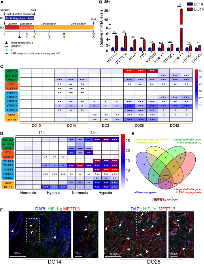
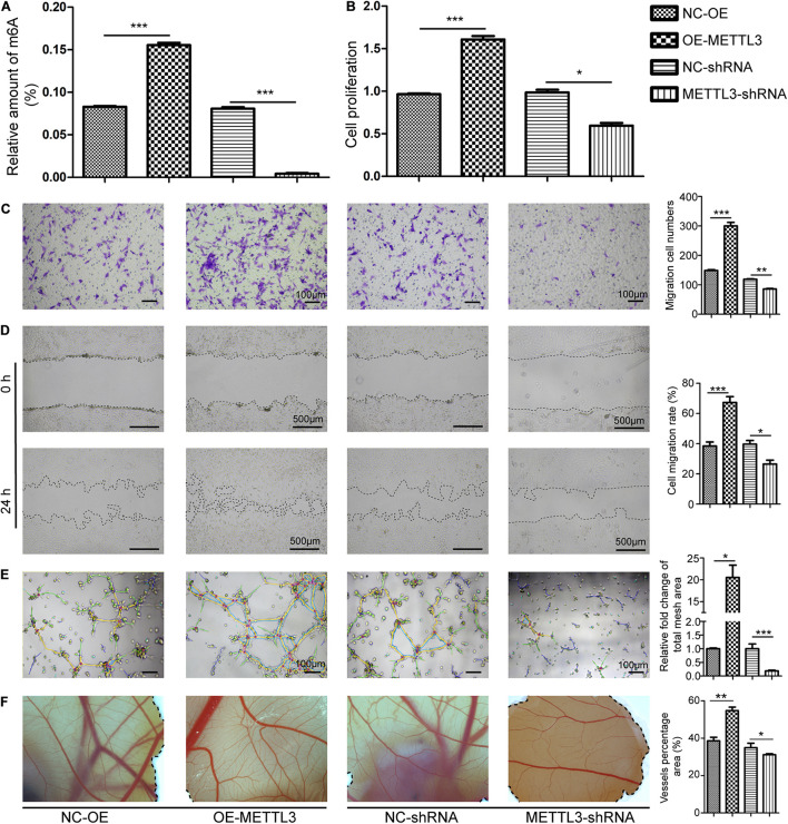
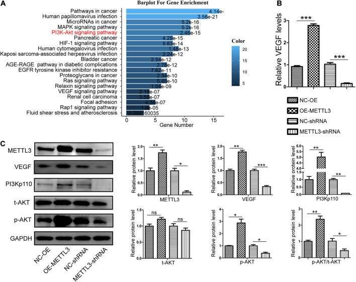
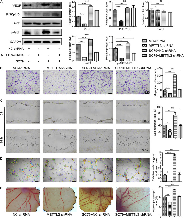
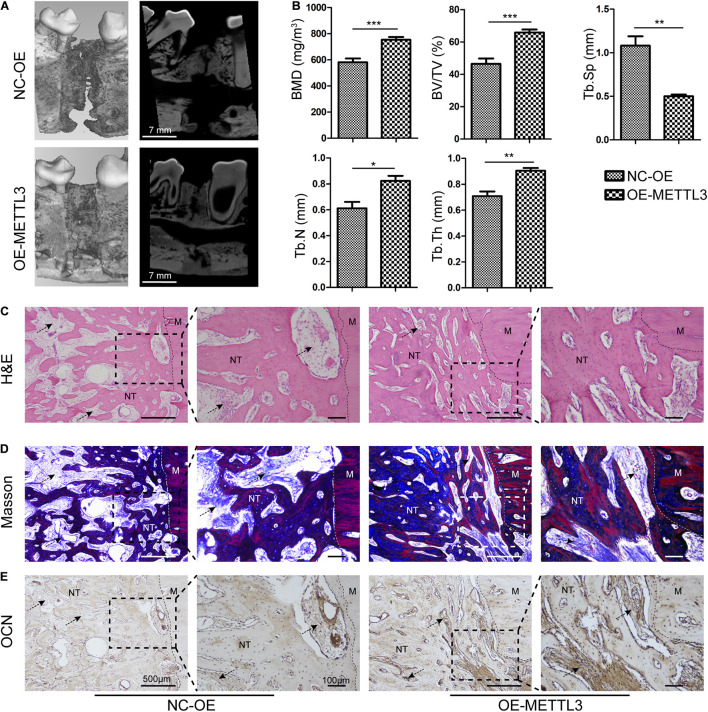
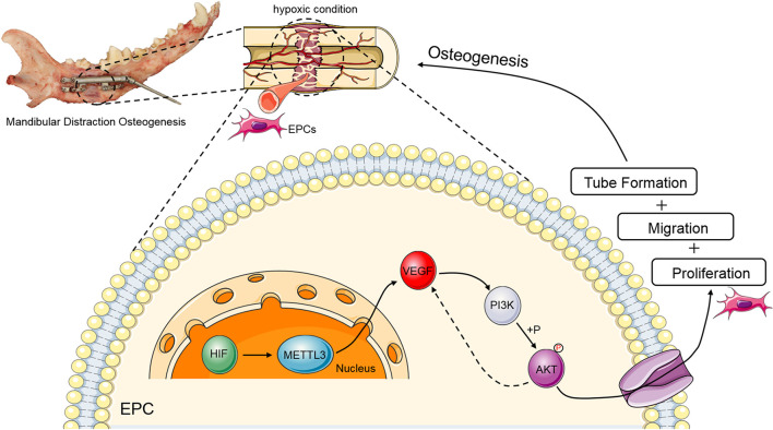
Distraction osteogenesis (DO) is used to treat large bone defects in the field of oral and maxillofacial surgery. Successful DO-mediated bone regeneration is dependent upon angiogenesis, and endothelial progenitor cells (EPCs) are key mediators of angiogenic processes. The N6-methyladenosine (m6A) methyltransferase has been identified as an important regulator of diverse biological processes, but its role in EPC-mediated angiogenesis during DO remains to be clarified. In the present study, we found that the level of m6A modification was significantly elevated during the process of DO and that it was also increased in the context of EPC angiogenesis under hypoxic conditions, which was characterized by increased METTL3 levels. After knocking down METTL3 in EPCs, m6A RNA methylation, proliferation, tube formation, migration, and chicken embryo chorioallantoic membrane (CAM) angiogenic activity were inhibited, whereas the opposite was observed upon the overexpression of METTL3. Mechanistically, METTL3 silencing reduced the levels of VEGF and PI3Kp110 as well as the phosphorylation of AKT, whereas METTL3 overexpression reduced these levels. SC79-mediated AKT phosphorylation was also able to restore the angiogenic capabilities of METTL3-deficient EPCs in vitro and ex vivo. In vivo, METTL3-overexpressing EPCs were additionally transplanted into the DO callus, significantly enhancing bone regeneration as evidenced by improved radiological and histological manifestations in a canine mandibular DO model after consolidation over a 4-week period. Overall, these results indicate that METTL3 accelerates bone regeneration during DO by enhancing EPC angiogenesis via the PI3K/AKT pathway.

Keywords: METTL3; PI3K/AKT signaling pathway; angiogenesis; distraction osteogenesis; endothelial progenitor cells.


Conflict of interest statement

The authors declare that the research was conducted in the absence of any commercial or financial relationships that could be construed as a potential conflict of interest.

Figures

FIGURE 1
FIGURE 1
Methyltransferase-like 3 (METTL3) expression is elevated in endothelial progenitor cell (EPC) angiogenic processes related to distraction osteogenesis (DO) regeneration. (A) Schematic overview of the canine DO and BF model employed in this study. (B) quantitative real-time polymerase chain reaction (qRT-PCR) results revealed that m6A methylation was increased in the DO callus tissue relative to the bone fracture (BF) model (n = 3 per group). (C) Heatmap of qRT-PCR results demonstrating the expression of N6-methyladenosine (m6A)-related genes during different stages of DO (n = 3/group). (D) Heatmap of qRT-PCR results in the context of EPC angiogenesis. Endothelial progenitor cells were cultured under hypoxic conditions in vitro to mimic the DO microenvironment. N6-methyladenosine and angiogenic cytokine expression levels were measured following culture for 24 h (n = 3/group). (E) Venn diagram demonstrating m6A-related gene overlap. METTL3 and YTHDC2 were specifically identified, and METTL3 was selected for further study owing to its high expression level. (F) IF staining for METTL3 and HIF-1α on DO14 and DO28. This analysis revealed that METTL3 was expressed on DO14 (white arrow) with gradual increases at later time points, and HIF-1α co-expression was also evident (n = 3/group). Scale bars: 500 and 100 μm. nsP > 0.05, *P < 0.05, **P < 0.01, ***P < 0.001. One-way ANOVA with Tukey’s post hoc test. Experiments were repeated in triplicate.
FIGURE 2
FIGURE 2
Methyltransferase-like 3 impacts m6A methylation and EPC-mediated angiogenesis. (A) Total m6A RNA methylation levels were quantified in EPCs following knockdown or overexpression. (B) Endothelial progenitor cell proliferation was measured via CCK-8 assay. METTL3 overexpression enhanced cellular proliferation, while silencing impairs such proliferation. (C–F) In vitro EPC angiogenesis was measured through Transwell (C), Wound healing (D), and Tube formation assays (E). Scale bars: 100 and 500 μm. (F) METTL3 affects ex vivo angiogenesis. CAM assays revealed that METTL3 overexpression significantly increased vascular density, while METTL3 inhibition had the opposite effect. NC-OE and OE-METTL3 groups or NC-shRNA and METTL3-shRNA groups were compared via Student’s t-tests. *P < 0.05, **P < 0.01, ***P < 0.001. All cell experiments were conducted in triplicates using three individual cell lines.
FIGURE 3
FIGURE 3
Methyltransferase-like 3 enhances PI3K/AKT signaling pathway activation and angiogenic cytokine production. (A) Gene Ontology enrichment analysis of the angiogenic targets of METTL3. (B) Following METTL3 knockdown or overexpression, the mRNA levels of the angiogenic cytokine VEGF were significantly altered. (C) PI3K/AKT signaling pathway and angiogenesis-related proteins including PI3Kp110, AKT, p-AKT, and VEGF were assessed via Western blotting in EPCs, with ImageJ being used for analysis. Analyses were conducted using data from three independent experiments. NC-OE and OE-METTL3 groups or NC-shRNA and METTL3-shRNA groups were compared via Student’s t-tests. nsP > 0.05, *P < 0.05, **P < 0.01, ***P < 0.001; n = 3 in each independent experiments.
FIGURE 4
FIGURE 4
Methyltransferase-like 3 regulates EPC angiogenesis via activating the PI3K/AKT pathway. (A) Western blotting analysis of PI3K/AKT pathway proteins in negative control and METTL3-deficient EPCs which were treated with SC79. (B–E) Migration assay (B,C), Tube formation (D), and CAM angiogenesis assays (E) were conducted using negative control and METTL3-deficient cells after SC79 treatment. Scale bars: 100 and 500 μm. These results indicated that injection with METTL3-knockdown EPCs was sufficient to reduce EPC angiogenic activity, whereas SC79 was able to reverse such impairment. Data are means ± SD and were compared via one-way ANOVA with Tukey’s post hoc test. nsP > 0.05, *P < 0.05, **P < 0.01, ***P < 0.001 vs. NC-shRNA; n = 3 per group, experiments were repeated in triplicate.
FIGURE 5
FIGURE 5
Methyltransferase-like 3 regulates angiogenesis and DO regeneration responses. (A,B) Representative 3D and longitudinal images (A) and quantitative analysis (B) of micro-CT data, including bone mineral density (BMD), bone volume/tissue volume (BV/TV), trabecular separation (Tb. Sp), trabecular number (Tb. N), and trabecular thickness (Tb. Th) of the mandibular distraction area after 4 weeks of consolidation. (C,D) For histological analyses, H&E (C) and Masson’s trichrome (D) staining of the MDO callus were performed in the different groups. (E) IHC assessment of OCN in the NC-OE and OE-METTL3 groups. NC-OE and OE-METTL3 groups were compared via Student’s t-tests. Arrows, extracellular matrix. M, Mandible, NT, Newly formed trabecula. Scale bars: 100 and 500 μm. *P < 0.05, **P < 0.01, ***P < 0.001 vs. NC-OE; n = 3 canine in each group.
FIGURE 6
FIGURE 6
Schematic overview of the mechanisms whereby METTL3 controls PI3K/AKT signaling activity in the context of DO-related EPC angiogenesis. During DO-related tissue regeneration, the DO gap is a hypoxic environment. Upon upregulation, METTL3 can target VEGF to gradually increase and positively regulate PI3K activity and AKT phosphorylation, with such PI3K/AKT signaling partially strengthening the expression of VEGF, ultimately promoting EPC proliferation, migration, and tube formation during DO and thereby facilitating osteogenesis.

References

    1. Atesok K., Li R., Stewart D. J., Schemitsch E. H. (2010). Endothelial progenitor cells promote fracture healing in a segmental bone defect model. J. Orthop. Res. 28 1007–1014. 10.1002/jor.21083 - DOI - PubMed
    1. Cetrulo C. L., Jr., Knox K. R., Brown D. J., Ashinoff R. L., Dobryansky M., Ceradini D. J., et al. (2005). Stem cells and distraction osteogenesis: endothelial progenitor cells home to the ischemic generate in activation and consolidation. Plast. Reconstr. Surg. 116 1053–1064; discussion 1065–1057. 10.1097/01.prs.0000178403.79051.70 - DOI - PubMed
    1. Choi I. H., Chung C. Y., Cho T.-J., Yoo W. J. (2002). Angiogenesis and mineralization during distraction osteogenesis. J. Korean Med. Sci. 17 435–447. 10.3346/jkms.2002.17.4.435 - DOI - PMC - PubMed
    1. Claesson-Welsh L., Welsh M. (2013). VEGFA and tumour angiogenesis. J. Intern. Med. 273 114–127. 10.1111/joim.12019 - DOI - PubMed
    1. Davidson E. H., Sultan S. M., Butala P., Tutela J. P., Canizares O., Wagner I. J., et al. (2011). Augmenting neovascularization accelerates distraction osteogenesis. Plast. Reconstr. Surg. 128 406–414. 10.1097/PRS.0b013e31821e6e2e - DOI - PubMed

LinkOut - more resources