Review
. 2021 Nov 1:8:737813.
doi: 10.3389/fmed.2021.737813. eCollection 2021.

Lichen Planus

Affiliations
Review

Lichen Planus

Katharina Boch et al. Front Med (Lausanne). .

Abstract

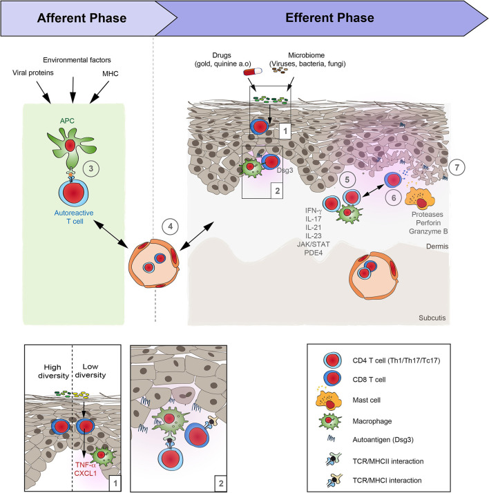
Lichen planus (LP) is a T cell-mediated disease affecting the stratified squamous epithelia of the skin and/or mucus membrane. Histologically, the disease is characterized by a lichenoid inflammatory infiltrate and vacuolar degeneration of the basal layer of the epidermis. LP has three major subtypes: Cutaneous, mucosal and appendageal LP. Rarely, it may affect the nails in the absence of skin and/or mucosal changes. LP may also be induced by several drugs, typically anti-hypertensive medication or be associated with infections, particularly viral hepatitis. The diagnosis is based on the clinical presentation and characteristic histological findings. Although the disease is often self-limiting, the intractable pruritus and painful mucosal erosions result in significant morbidity. The current first-line treatment are topical and/or systemic corticosteroids. In addition, immunosuppressants may be used as corticosteroid-sparing agents. These, however are often not sufficient to control disease. Janus kinase inhibitors and biologics (anti-IL-12/23, anti-IL17) have emerged as novel future treatment options. Thus, one may expect a dramatic change of the treatment landscape of LP in the near future.

Keywords: T-cell mediated; inflammation; lichen planus; skin disease; treatment.


Conflict of interest statement

The authors declare that the research was conducted in the absence of any commercial or financial relationships that could be construed as a potential conflict of interest.

Figures

Figure 1
Figure 1
Clinical and histological hallmarks of lichen planus. (a–f) Cutaneous lichen planus (LP). (a) Polygonal, flat-topped, violaceous confuting plaques with fine white scales on the inner wrist in a patient with localized LP. (b) Symmetric red plaques on the back of a patient with generalized cutaneous LP. (c) Thick reddish-brown plaques on the arms of a patient with hypertrophic LP. (e) Blister on the 3rd toe along with widespread red plaques with whit streaks in a patient with lichen planus pemphigoides. (f) Linear lichen planus. (g) Direct immunofluorescence microcopy staining with fibrin deposition in the epidermis (400 ×). (h) The histology from a skin biopsy from a lichen planus lesion characteristically shows an irregularly epidermis with saw-toothed rete ridges, hypergranulosis, liquefaction degeneration of the dermal-epidermal junction and a lichenoid (band-like) lymphocytic infiltrate (H&E staining, 200 ×). (i–l) Appendageal LP. (i) Scaring alopecia and inflammation around hair follicles along the frontal scalp hair margin in a patient with frontal fibrosing alopecia. (j) Image from a patent with lichen planopilaris. (k) Lichenoid interface dermatitis of the hair infundibulum and apoptotic keratinocytes (Civatte bodies) and fibrous tracts in a biopsy from a patient with frontal fibrosing alopecia. (l) Grooved and ridged nails in a patient with nail LP. (m–s) Mucosal LP. (m) Wickham striae in the oral mucosa of a patient with oral LP. (n) Severe ulcera of the tounge in a patient with erosive oral LP. (o) Parakeratosis, acanthosis. band of inflammatory cells just beneath the epidermis, plasma cells in infiltrate in an oral biopsy from a patient with oral LP. (p) Severe vulval ulcerations in a patient with vulval LP. (q) Erythema and erosions in a patient with vulval LP. (r) Wickham striae on the glans penis in a patient with penile LP. (s) Occasional parakeratosis, irregularly thickened epidermis, apoptotic basal keratinocytes, lymphohistiocytic infiltrate in a biopsy from a patient with genital LP.
Figure 2
Figure 2
Schematic overview of lichen planus pathogenesis. Based on the increasing evidence of the autoimmune nature of lichen planus (LP), its pathogenesis may be divided in two distinct phases: Afferent phase, where tolerance to autoantigens is lost, and the efferent phase that is characterized by a T cell-driven skin inflammation. (1) Under steady state conditions the diversity of the cutaneous microbiome can alter the “inflammatory state” of the skin. If the diversity of the bacterial communities on the skin is low, this is associated with an increased expression of pro-inflammatory cytokines such as TNFα and CXCL1, as well as an increase of CD11c, suggesting an increased presence of macrophages. (2, 3) Following this presumed injury, the loss of tolerance in LP occurs in the context of an association with the MHC that may additionally be shaped by associated viral infections and other environmental factors. The site of the APC/T-cell interaction is so far unknown. It may occur locally and/or in skin draining lymph nodes. (4) Next, autoreactive T cells reach the skin by extravasation from the blood vessels. (5) Within the skin, T cells become activated by binding to the specific autoantigens. Effector functions of cytotoxic T cells is mediated by (6) proteases, such as granzyme B, as well as perforin. In addition, (7) mast cells become activated and may further aggravate inflammation in LP. However, the precise sequence of events and their interactions are only incompletely understood.

Similar articles

Cited by

References

    1. Boyd AS, Neldner KH. Lichen planus. J Am Acad Dermatol. (1991) 25:593–619. 10.1016/0190-9622(91)70241-S - DOI - PubMed
    1. Shiohara T, Moriya N, Mochizuki T, Nagashima M. Lichenoid tissue reaction (LTR) induced by local transfer of Ia-reactive T-cell clones. II LTR by epidermal invasion of cytotoxic lymphokine-producing autoreactive T cells. J Investig Dermatol. (1987) 89:8–14. 10.1111/1523-1747.ep12523539 - DOI - PubMed
    1. Sugerman PB, Satterwhite K, Bigby M. Autocytotoxic T-cell clones in lichen planus. Br J Dermatol. (2000) 142:449–56. 10.1046/j.1365-2133.2000.03355.x - DOI - PubMed
    1. Shiohara T, Moriya N, Nagashima M. Induction and control of lichenoid tissue reactions. Springer Semin Immunopathol. (1992) 13:369–85. 10.1007/BF00200535 - DOI - PubMed
    1. Yasukawa M, Ohminami H, Arai J, Kasahara Y, Ishida Y, Fujita S. Granule exocytosis, and not the fas/fas ligand system, is the main pathway of cytotoxicity mediated by alloantigen-specific CD4(+) as well as CD8(+) cytotoxic T lymphocytes in humans. Blood. (2000) 95:2352–5. 10.1182/blood.V95.7.2352 - DOI - PubMed

LinkOut - more resources