. 2021 Oct;9(20):1575.
doi: 10.21037/atm-21-5066.

Synergistic effect of chidamide and venetoclax on apoptosis in acute myeloid leukemia cells and its mechanism

Affiliations

Synergistic effect of chidamide and venetoclax on apoptosis in acute myeloid leukemia cells and its mechanism

Gangping Li et al. Ann Transl Med. 2021 Oct.

Abstract

Background: Acute myeloid leukemia (AML) is a hematological malignancy with a low remission rate and high recurrence rate. Overexpression of the antiapoptotic protein Bcl-2 is associated with a lower overall survival rate in AML patients. Venetoclax (ABT199) is a selective inhibitor of Bcl-2 that has a significant effect in AML, but single-drug resistance often occurs due to the high expression of Mcl-1 protein. Studies have confirmed that chidamide can downregulate the expression levels of Bcl-2 and Mcl-1 and induce apoptosis.

Methods: This study aimed to use AML cell lines and primary cells to study the effects of venetoclax and chidamide combination therapy on AML cell apoptosis, the cell cycle, and changes in related signaling pathways in vitro; establish an AML mouse model to observe the efficacy and survival time of combination therapy in vivo; and analyze the drug effects with multi-omics sequencing technology. The changes in gene and protein expression before and after treatment were examined to clarify the molecular mechanism driving the synergistic effect of the two drugs.

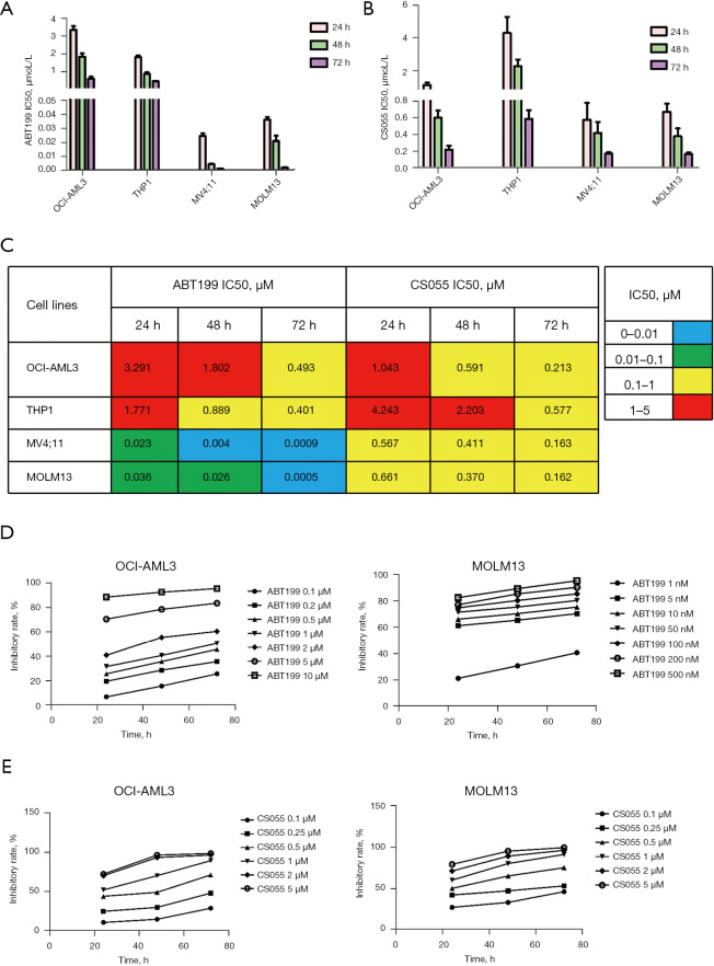
Results: (I) Both venetoclax and chidamide promoted apoptosis in AML cell lines and primary cells in a time- and concentration-dependent manner. The effect was further enhanced when the two drugs were combined, and a synergistic effect was observed (combination index <1). (II) At both the mRNA and protein levels, the expression of Mcl-1 was upregulated by venetoclax and downregulated by chidamide, and the expression of Mcl-1 decreased further after combination treatment. (III) Transcriptome sequencing showed that differentially expressed genes in the combination group compared with the venetoclax monotherapy group were mainly enriched in the PI3K-AKT pathway and JAK2/STAT3 pathway. Moreover, qRT-PCR and Western blot confirmed these results. (IV) The combination therapy group exhibited significantly inhibited disease progression and a prolonged survival time among AML mice.

Conclusions: Chidamide combined with venetoclax synergistically promoted apoptosis in AML cell lines and primary cells by inhibiting activation of the PI3K/AKT pathway and JAK2/STAT3 pathway.

Keywords: Chidamide; acute myeloid leukemia (AML); apoptosis; mechanism; venetoclax.


Conflict of interest statement

Conflicts of Interest: All authors have completed the ICMJE uniform disclosure form (available at https://dx.doi.org/10.21037/atm-21-5066). The authors have no conflicts of interest to declare.

Figures

Figure 1
Figure 1
The IC50 values of ABT199 (venetoclax) and CS055 (chidamide) on AML cell lines (OCI-AML3, THP-1, MV4;11, and MOLM13) at 24, 48 and 72 h, and the proliferation inhibition curves of different concentrations of ABT199 and CS055 on OCI-AML3 and MOLM13 cell lines at 24, 48 and 72 h. (A) IC50 values of four AML cell lines treated with ABT199 for 24, 48, and 72 h. (B) IC50 values of four AML cell lines treated with CS055 for 24, 48, and 72 h. (C) IC50 values of the two drugs in four AML cell lines at 24, 48, and 72 h. (D) The proliferation inhibition curve of ABT199 for OCI-AML3 and MOLM13 at 24, 48, and 72 h. (E) The proliferation inhibition curve of CS055 for OCI-AML3 and MOLM13 at 24, 48, and 72 h. AML, acute myeloid leukemia.
Figure 2
Figure 2
ABT199 (Venetoclax) and CS055(chidamide) single and combined effects on the apoptosis rate of AML cell line OCI-AML3 and MOLM13 at 8 and 24 h, and the changes of apoptosis related genes and proteins at 24 h. (A) Apoptosis histograms of the OCI-AML3 treated with ABT199 or CS055 alone or in combination for 8 and 24 h. (B) Apoptosis histograms of the OCI-AML3 treated with ABT199 or CS055 alone or in combination for 8 and 24 h. (C) ABT199 and CS055 monotherapy and combination treatment of primary AML cells. (D) Changes in apoptosis-related proteins detected by qRT-PCR. (E) Changes in apoptosis-related proteins detected by Western blot. (F) Changes in Bcl-2 family apoptotic regulatory proteins detected by Western blot. *, P<0.05; **, P<0.01; ***, P<0.001, compared with the control group. #, P<0.05; ##, P<0.01; ###, P<0.001. AML, acute myeloid leukemia.
Figure 3
Figure 3
Histogram of changes in the mitochondrial membrane potential in the OCI-AML3, THP1, MV4;11, and MOLM13 cell lines treated with ABT199 (venetoclax) and CS055 (chidamide). *, P<0.05; ***, P<0.001, compared with the control group.
Figure 4
Figure 4
Cell cycle histogram, cycle related genes and protein changes of AML cell lines (OCI-AML3, MOLM13) and primary cells treated with ABT199 (venetoclax) and CS055 (chidamide) for 24 h. (A) Cell cycle analysis after ABT199 and CS055 monotherapy and combination therapy in different AML cell lines and primary AML cells with flow cytometry. (B) Changes in cell cycle-related genes before and after treatment, as detected by qRT-PCR. The data were statistically analyzed with a t-test. (C) Changes in cell cycle-related proteins before and after treatment, as detected by Western blot. *, P<0.05; **, P<0.01; ***, P<0.001, compared with the control group. #, P<0.05; ##, P<0.01; ###, P<0.001. AML, acute myeloid leukemia.
Figure 5
Figure 5
Differential expression gene analysis and KEGG pathway enrichment results of transcriptome sequencing samples, as well as relevant results of signal pathway verification by qRT-PCR and Western blot. (A) Volcano map of differentially expressed genes. (B) Genes with the most obvious differences in expression. (C) Enrichment analysis of KEGG pathways. (D) mRNA levels of AKT, HDAC1, SOCS3, JAK2, and STAT3 expressed in OCI-AML, MOLM13, and primary cells treated with ABT199 (venetoclax) and CS055 (chidamide) for 24 h, as analyzed by qRT-PCR. The data were statistically analyzed by a t-test. (E) PI3K-AKT and JAK2/STAT3 signaling pathway-related proteins and HDAC1 protein expression levels in the OCI-AML3 and MOLM13 cell lines and AML primary cells after 24 h of treatment with ABT199 and CS055. *, P<0.05; **, P<0.01; ***, P<0.001, compared with the control group. #, P<0.05; ##, P<0.01; ###, P<0.001. AML, acute myeloid leukemia.
Figure 6
Figure 6
Signaling pathway of ABT199 (venetoclax) combined with CS055 (chidamide) in AML cells. AML, acute myeloid leukemia.
Figure 7
Figure 7
The effects of ABT199 (venetoclax) and CS055 (chidamide) on AML xenotransplantation mice, and the negative group was used as the control. (A) Survival curve of AML xenograft mice treated with ABT199 and CS055 alone or in combination. (B) Fluorescence intensity of AML xenograft mice in the monotherapy groups and the combination therapy group. (C) Tumorigenesis of AML xenograft mice treated with ABT199 and CS055 alone or in combination. (D) On the 10th day of administration, three mice in each group were sacrificed for in vivo imaging. (E) The number of CD45+CD33+ cells in the bone marrow of AML xenotransplantation mice treated with ABT199 and CS055 alone or in combination for 10 days. (F) Body weight curve of mice with AML xenotransplantation treated with ABT199 and CS055 for 14 days. (G) Ten days later, mice were sacrificed, and the liver, spleen, and lung were stained with HE (magnification, ×200 for liver and lung, ×400 for spleen). (H) On the 10th day, the mice were sacrificed, and the liver, spleen and lung were stained for hCD45+ with immunohistochemistry (magnification: ×200 for liver and lung, ×400 for spleen). A t-test was used to analyze the data. *, P<0.05; **, P<0.01; ***, P<0.001. ns, not significant; AML, acute myeloid leukemia.

Similar articles

Cited by

References

    1. Dombret H, Gardin C. An update of current treatments for adult acute myeloid leukemia. Blood 2016;127:53-61. 10.1182/blood-2015-08-604520 - DOI - PMC - PubMed
    1. Tamamyan G, Kadia T, Ravandi F, et al. Frontline treatment of acute myeloid leukemia in adults. Crit Rev Oncol Hematol 2017;110:20-34. 10.1016/j.critrevonc.2016.12.004 - DOI - PMC - PubMed
    1. Bose P, Vachhani P, Cortes JE. Treatment of Relapsed/Refractory Acute Myeloid Leukemia. Curr Treat Options Oncol 2017;18:17. 10.1007/s11864-017-0456-2 - DOI - PubMed
    1. Konopleva M, Pollyea DA, Potluri J, et al. Efficacy and Biological Correlates of Response in a Phase II Study of Venetoclax Monotherapy in Patients with Acute Myelogenous Leukemia. Cancer Discov 2016;6:1106-17. 10.1158/2159-8290.CD-16-0313 - DOI - PMC - PubMed
    1. DiNardo CD, Rausch CR, Benton C, et al. Clinical experience with the BCL2-inhibitor venetoclax in combination therapy for relapsed and refractory acute myeloid leukemia and related myeloid malignancies. Am J Hematol 2018;93:401-7. 10.1002/ajh.25000 - DOI - PubMed