Review
. 2022 Apr 18;145(2):426-440.
doi: 10.1093/brain/awab419.

The neuroradiology of progressive multifocal leukoencephalopathy: a clinical trial perspective

Affiliations
Review

The neuroradiology of progressive multifocal leukoencephalopathy: a clinical trial perspective

Laura E Baldassari et al. Brain. .

Abstract

Progressive multifocal leukoencephalopathy (PML) is an opportunistic infection of the CNS caused by the JC virus, which infects white and grey matter cells and leads to irreversible demyelination and neuroaxonal damage. Brain MRI, in addition to the clinical presentation and demonstration of JC virus DNA either in the CSF or by histopathology, is an important tool in the detection of PML. In clinical practice, standard MRI pulse sequences are utilized for screening, diagnosis and monitoring of PML, but validated imaging-based outcome measures for use in prospective, interventional clinical trials for PML have yet to be established. We review the existing literature regarding the use of MRI and PET in PML and discuss the implications of PML histopathology for neuroradiology. MRI not only demonstrates the localization and extent of PML lesions, but also mirrors the tissue destruction, ongoing viral spread, and resulting inflammation. Finally, we explore the potential for imaging measures to serve as an outcome in PML clinical trials and provide recommendations for current and future imaging outcome measure development in this area.

Keywords: clinical trial design; imaging outcome measures; magnetic resonance imaging; progressive multifocal leukoencephalopathy.


Figures

Figure 1
Figure 1
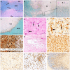
PML pathology. Two demyelinated PML lesions located at the grey-white matter junction are indicated by arrows [initial lesions shown in A, Luxol fast blue/periodic acid Schiff (LFB/PAS) staining]. Lesions may enlarge either by extension (not shown) or expansion due to viral progression at the lesion edge [B; LFB/PAS (blue/pink) plus anti-VP1 (viral protein 1) (brown)]. Numerous virally infected cells are found at the lesion edge, as shown in the inset. Finally, the lesions fuse to form large demyelinated areas (pale regions marked with asterisk in B) and band-like demyelination at the border between the cortex and white matter (C; LFB/PAS plus anti-VP1). Typical histological features include virally infected glial cells with ground glass oligodendrocytes (D; haematoxylin and eosin staining; ground glass oligodendrocytes are indicated by arrows) and bizarre, pleomorphic astrocytes [E; anti-glial fibrillary acidic protein (GFAP); astrocytes are indicated by arrows]. Lesions may be destructive, leading to a pronounced axonal damage in the lesion centre (F; Bielschowsky silver stain) and even necrotic lesions (G; haematoxylin and eosin). The inflammatory infiltrate within PML lesions consists of numerous macrophages (H; KiM1P staining) and typically few lymphocytes (I; anti-CD3 staining), due to immunosuppression. In contrast, numerous lymphocytes are found with immune reconstitution (J; anti-CD3). JCV infection may also cause granule cell neuronopathy (K; Pab 2003 staining for large T-antigen). Scale bars = 1 mm in A–C; 50 μm in D and E; 20 μm in G and H; 100 μm in F and I–K. Counterstain in E and HK: haematoxylin. Ctx = cortex; NAWM = normal-appearing white matter; WM = white matter.
Figure 2
Figure 2
Histopathological correlations of MRI findings in classic and inflammatory PML. Brain MRI and corresponding histopathology (from different cases) of classic PML (top row) and inflammatory PML (bottom row). Histopathology for both classic and inflammatory PML is characterized by positive SV40 T antigen staining, indicating JCV infection. However, inflammatory PML lesions typically show lower numbers of infected cells. In classic PML, few CD8+ T cells and CD138+ plasma cells are present, whereas in inflammatory PML, there is pronounced infiltration of CD8+ T cells and CD138+ plasma cells. T2-FLAIR images show the extent of the PML lesion. T2-weighted turbo/fast spin echo sequences may show intralesional vacuoles (bottom row). In early stages, the T1-weighted signal intensity can be normal (bottom row), while in advanced stages, it is low (top row). In inflammatory PML, contrast-enhanced T1-weighted images often show enhancement (bottom row). High signal intensity on DWI in classic PML represents areas of active inflammation and viral replication leading to swelling of oligodendrocytes. Lesions show heterogenous ADC values, but rarely is diffusion markedly restricted. Scale bars = 50 µm.
Figure 3
Figure 3
Imaging findings of PML on multisequence MRI. Axial brain MRI images of a patient presenting with PML lesions in bilateral occipital lobes, primarily in the subcortical white matter and adjacent cortical grey matter. These images are consistent with a classic MRI pattern of PML, with high signal intensity on T2-weighted images and DWI, and low signal intensity on the contrast-enhanced T1-weighted images (arrows). On SWI, bands of low signal intensity adjacent to cortical grey matter can be observed, suggesting deposition of paramagnetic compounds such as iron (arrows).
Figure 4
Figure 4
Axial T2-weighted and contrast-enhanced T1-weighted MRI of a PML patient treated with BK virus-specific allogeneic T-cells. This patient’s MRIs demonstrated lesion growth and contrast enhancement at early stages (progressing) but stability and/or retraction, with resolution of contrast enhancement, weeks after treatment initiation.

References

    1. Cortese I, Reich DS, Nath A. Progressive multifocal leukoencephalopathy and the spectrum of JC virus-related disease. Nat Rev Neurol. 2020;17(1):37. - PMC - PubMed
    1. Brew BJ, Davies NW, Cinque P, Clifford DB, Nath A. Progressive multifocal leukoencephalopathy and other forms of JC virus disease. Nat Rev Neurol. 2010;6(12):667–679. - PubMed
    1. Bowen LN, Smith B, Reich D, Quezado M, Nath A. HIV-associated opportunistic CNS infections: Pathophysiology, diagnosis and treatment. Nat Rev Neurol. 2016;12(11):662–674. - PubMed
    1. Major EO, Yousry TA, Clifford DB. Pathogenesis of progressive multifocal leukoencephalopathy and risks associated with treatments for multiple sclerosis: A decade of lessons learned. Lancet Neurol. 2018;17(5):467–480. - PubMed
    1. Wijburg MT, Warnke C, McGuigan C, et al. Pharmacovigilance during treatment of multiple sclerosis: Early recognition of CNS complications. J Neurol Neurosurg Psychiatry. 2021;92(2):177–188. - PubMed

MeSH terms