

Eye-Transcriptome and Genome-Wide Sequencing for Scolecophidia: Implications for Inferring the Visual System of the Ancestral Snake
- PMID: 34791190
- PMCID: PMC8643396
- DOI: 10.1093/gbe/evab253
Eye-Transcriptome and Genome-Wide Sequencing for Scolecophidia: Implications for Inferring the Visual System of the Ancestral Snake
Abstract
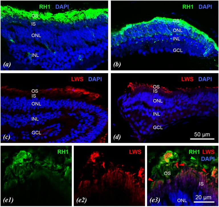
Molecular genetic data have recently been incorporated in attempts to reconstruct the ecology of the ancestral snake, though this has been limited by a paucity of data for one of the two main extant snake taxa, the highly fossorial Scolecophidia. Here we present and analyze vision genes from the first eye-transcriptomic and genome-wide data for Scolecophidia, for Anilios bicolor, and A. bituberculatus, respectively. We also present immunohistochemistry data for retinal anatomy and visual opsin-gene expression in Anilios. Analyzed in the context of 19 lepidosaurian genomes and 12 eye transcriptomes, the new genome-wide and transcriptomic data provide evidence for a much more reduced visual system in Anilios than in non-scolecophidian (=alethinophidian) snakes and in lizards. In Anilios, there is no evidence of the presence of 7 of the 12 genes associated with alethinophidian photopic (cone) phototransduction. This indicates extensive gene loss and many of these candidate gene losses occur also in highly fossorial mammals with reduced vision. Although recent phylogenetic studies have found evidence for scolecophidian paraphyly, the loss in Anilios of visual genes that are present in alethinophidians implies that the ancestral snake had a better-developed visual system than is known for any extant scolecophidian.
Keywords: Squamata; gene loss; opsins; phylogeny; regressive evolution; vision.
© The Author(s) 2021. Published by Oxford University Press on behalf of the Society for Molecular Biology and Evolution.
Figures


References
-
- Bellairs ADA. 1972. Comments on the evolution and affinities of snakes. In: Joysey KA, Kemp TS, editors.Studies in vertebrate evolution. Edinburgh: Oliver & Boyd. p. 157–172.
-
- Bellairs ADA, Underwood G.. 1951. The origin of snakes. Biol Rev Camb Philos Soc. 26(2):193–237. - PubMed
-
- Bhattacharyya N, Darren B, Schott RK, Tropepe V, Chang BSW.. 2017. Cone-like rhodopsin expressed in the all-cone retina of the colubrid pine snake as a potential adaptation to diurnality. J Exp Biol. 220(Pt 13):2418–2425. - PubMed
Publication types
MeSH terms
Substances
LinkOut - more resources
Full Text Sources
