. 2022 Jan 7;375(6576):86-91.
doi: 10.1126/science.abl4732. Epub 2021 Nov 18.

Cryo-EM structure of human GPR158 receptor coupled to the RGS7-Gβ5 signaling complex

Affiliations

Cryo-EM structure of human GPR158 receptor coupled to the RGS7-Gβ5 signaling complex

Dipak N Patil et al. Science. .

Abstract

GPR158 is an orphan G protein–coupled receptor (GPCR) highly expressed in the brain, where it controls synapse formation and function. GPR158 has also been implicated in depression, carcinogenesis, and cognition. However, the structural organization and signaling mechanisms of GPR158 are largely unknown. We used single-particle cryo–electron microscopy (cryo-EM) to determine the structures of human GPR158 alone and bound to an RGS signaling complex. The structures reveal a homodimeric organization stabilized by a pair of phospholipids and the presence of an extracellular Cache domain, an unusual ligand-binding domain in GPCRs. We further demonstrate the structural basis of GPR158 coupling to RGS7-Gβ5. Together, these results provide insights into the unusual biology of orphan receptors and the formation of GPCR-RGS complexes.


Conflict of interest statement

Competing interests: Authors declare that they have no competing interests.

Figures

Fig. 1.
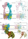
Fig. 1.. CryoEM structures of GPR158 in its apo and RGS7-Gβ5 bound states.
(A) CryoEM map (left) and model (right) of GPR158 homodimer in ribbon representation with protomers colored in cyan and pink. Phospholipids PE and PI, and cholesterols are shown in green, blue, and yellow colors, respectively. (B) CryoEM map (left) and model (right) of GPR158 homodimer complexed with RGS7-Gβ5 in ribbon representation colored as (A) and RGS7 and Gβ5 are shown in deep-olive and brown color.
Fig. 2.
Fig. 2.. Organization of GPR158 transmembrane domain and its homodimer interface.
(A) Overall arrangement of the 7TM region of GPR158 protomers is shown as side and top view. Phospholipids PE and PI are identified at the cavity formed by TM dimeric interface are shown in spheres representation. 7TM protomers and phospholipids are colored as Fig.1. (B) Close-up view of the extracellular loop region. ECL2 caps the extracellular pocket by interacting with TM3, ECL1, and ECL3 residues. ECL2 C573 preserves the conserved disulfide bond with TM3 C481. ECL2 is also stabilized by interaction with a stalk-TM linker that connects ectodomain with 7TM. (C) The 7TM dimer interface is formed at two sites (I and II), the extra- and intracellular side. Direct contacts at the extracellular side are formed by TM4, TM5, and ECL2 of both protomers, and contacts are shown in the right panels. Intracellular side interface is formed by TM3 and ICL2 of both protomers and contacts formed by ICL2 are shown in the right panel.
Figure 3.
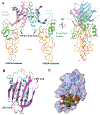
Figure 3.. A unique organization of the GPR158 ectodomain featuring the Cache domain.
(A) Side view of GPR158 ectodomain consisting of N-terminal domain (NTD), Cache domain, and stalk region. The GPR158 ectodomain forms a dimeric interface with the Cache domain. (B) The Cache domain is composed of six antiparallel β-sheets flanked by α-helices. The density for α3 helix is not well resolved and is represented as dotted cylinder at respective position. The missing flexible loops in the model are shown in dotted lines. (C) Cache domain putative ligand-binding pocket. The curved sheets form a putative ligand-binding pocket (dotted oval), equivalent to the prokaryotes extracellular Cache domain ligand binding site, generates an amphipathic environment. The putative ligand interacting residues shown in brown color. However, densities for the most side chains of pocket residues are not well resolved and have high B-factor. The putative ligand binding pocket is possibly capped by dynamic α3 helix from one side.
Figure 4.
Figure 4.. Mechanism of GPR158 interaction with RGS7.
(A) RGS7 forms two distinct binding sites (I and II) on GPR158, both created by the dimerization of GPR158. The first binding interface is formed between GPR158 CT-CC and RGS7 DEP-DHEX domain. The second interface is formed by GPR158 7TM and RGS7 DHEX domain. In addition, β-hairpin loop of RGS7 is inserted into the membrane (shown as III) and could facilitate the orientation of RGS7 towards the membrane. (B and C) Conformational rearrangement on GPR158 TM dimeric interface and RGS7 upon complex formation. The TM3 of one protomer shifts toward the 7TM core while dissociating it from another protomer to accommodate RGS7 at the interface (B). Large conformation shift at Eα1Eα2 loop of RGS7 DHEX domain along with rearrangement of β-hairpin loop and helices that shifted up towards the membrane (C).

References

    1. Hauser AS et al. Pharmacogenomics of GPCR Drug Targets. Cell 172, 41–54 e19 (2018). - PMC - PubMed
    1. Sriram K, Insel PA, G Protein-Coupled Receptors as Targets for Approved Drugs: How Many Targets and How Many Drugs? Mol Pharmacol 93, 251–258 (2018). - PMC - PubMed
    1. Gilman AG, G proteins: transducers of receptor-generated signals. Annu Rev Biochem 56, 615–649 (1987). - PubMed
    1. Shukla AK, Xiao K, Lefkowitz RJ, Emerging paradigms of beta-arrestin-dependent seven transmembrane receptor signaling. Trends Biochem Sci 36, 457–469 (2011). - PMC - PubMed
    1. Hollinger S, Hepler JR, Cellular regulation of RGS proteins: modulators and integrators of G protein signaling. Pharmacol Rev 54, 527–559 (2002). - PubMed

Publication types

MeSH terms