. 2021 Nov 12;18(sup2):804-817.
doi: 10.1080/15476286.2021.1999103. Epub 2021 Nov 18.

Nsp1 of SARS-CoV-2 stimulates host translation termination

Affiliations

Nsp1 of SARS-CoV-2 stimulates host translation termination

Alexey Shuvalov et al. RNA Biol. .

Abstract

Nsp1 of SARS-CoV-2 regulates the translation of host and viral mRNAs in cells. Nsp1 inhibits host translation initiation by occluding the entry channel of the 40S ribosome subunit. The structural study of the Nsp1-ribosomal complexes reported post-termination 80S complex containing Nsp1, eRF1 and ABCE1. Considering the presence of Nsp1 in the post-termination 80S ribosomal complex, we hypothesized that Nsp1 may be involved in translation termination. Using a cell-free translation system and reconstituted in vitro translation system, we show that Nsp1 stimulates peptide release and formation of termination complexes. Detailed analysis of Nsp1 activity during translation termination stages reveals that Nsp1 facilitates stop codon recognition. We demonstrate that Nsp1 stimulation targets eRF1 and does not affect eRF3. Moreover, Nsp1 increases amount of the termination complexes at all three stop codons. The activity of Nsp1 in translation termination is provided by its N-terminal domain and the minimal required part of eRF1 is NM domain. We assume that the biological meaning of Nsp1 activity in translation termination is binding with the 80S ribosomes translating host mRNAs and remove them from the pool of the active ribosomes.

Keywords: Nsp1; SARS-CoV-2; eRF1; eRF3; ribosome.


Conflict of interest statement

No potential conflict of interest was reported by the author(s).

Figures

Figure 1.
Figure 1.
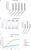
Nsp1 activates peptidyl-tRNA hydrolysis with the release factors. (A) In vitro Nluc mRNA translation in RRL in the presence/absence of Nsp1. (B) Termi-Luc peptide release assay in the presence/absence of Nsp1 and the release factors. GST was added to the reaction as a negative control. Time progress curves showing luminescence (in relative luminescence units, rlu) with NLuc released from the ribosome complex upon treatment with the proteins of interest, number of repeats, n = 3, mean±SD
Figure 2.
Figure 2.
Nsp1 increases TC formation. Examples of raw toe-printing data and relative quantitative analysis of the TC formation efficiency induced by the release factors in the presence of Nsp1 or GST as a negative control. TC formation efficiency induced by release factors was taken as 1. (A) Toe-print analysis of TCs formed by addition to the preTCs of eRF1 and Nsp1. (B) Toe-print analysis of TCs formed by addition to the preTCs of eRF1(AGQ) and Nsp1. (C) Toe-print analysis of TCs formed by addition to the preTCs of eRF1+eRF3a+GTP and Nsp1. (D) Toe-print analysis of TCs formed by addition to the preTCs of eRF1(AGQ)+eRF3a+GTP and Nsp1. r.u. – relative units. Positions of preTCs and TCs are labelled by white and black triangles, respectively. Red stars indicate the increased quantity of ribosomal complexes, shifted from the preTC to the TC state. The error bars represent the standard error of mean, stars (**) mark a significant difference from the respective control P < 0.01 (n = 3)
Figure 3.
Figure 3.
Nsp1 stimulates translation termination independently of GTP hydrolysis. (A) GTPase activity of eRF3a in the presence of the ribosome subunits, eRF1 and Nsp1, number of repeats, n = 4, mean±SE. (B) Nsp1 stimulation of TC formation does not depend on GTP hydrolysis. TC formation was induced by the addition to preTC of eRF1+eRF3a+GDPCP and Nsp1 or GST as a negative control. TC formation efficiency induced by eRF1+eRF3a+GDPCP was taken as 1. The TC corresponds to the black triangle, and the preTC corresponds to the white triangle, r.u. – relative units. Red star indicates the increased quantity of ribosomal complexes, shifted from the preTC to the TC state. The error bars represent the standard error of mean, stars (**) mark a significant difference from the respective control P < 0.01 (n = 3). (C) Termi-Luc peptide release assay in the presence/absence of Nsp1 and NM-eRF1 or eRF1. Time progress curves showing luminescence (in relative luminescence units, rlu) with NLuc released from the ribosome complex upon treatment with the proteins of interest, number of repeats, n = 3, mean±SD
Figure 4.
Figure 4.
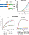
Activity of the Nsp1 mutants in translation termination. (A) Scheme of the Nsp1 forms used in the experiments. (B) In vitro Nluc mRNA translation in RRL in the presence/absence of Nsp1 mutants. (C) Termi-Luc peptide release assay in the presence/absence of Nsp1 mutants and the release factors. Time progress curves showing luminescence (in relative luminescence units, rlu), number of repeats, n = 3, mean±SD
Figure 5.
Figure 5.
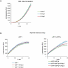
Activity of the Nsp1 domains in translation termination. (А) In vitro Nluc mRNA translation in RRL in the presence/absence of N or C domains of Nsp1, n = 3, mean±SD. (B) Termi-Luc peptide release assay in the presence/absence of N or C domains of Nsp1 and the release factors. Time progress curves showing luminescence (in relative luminescence units, rlu) with NLuc released from the ribosome complex upon treatment with the proteins of interest, n = 3, mean±SD
Figure 6.
Figure 6.
NSP1 increases TC formation at all three stop codons. (А) Toe-print analysis of the TCs formed by addition to the preTCs of eRF1 and Nsp1 and relative quantitative analysis of the TC formation efficiency at different stop codons (UAA, UAG, UGA) in the presence of eRF1 and Nsp1. TC formation efficiency induced by eRF1 alone was taken as 1. (B) Toe-print analysis of TCs formed by addition to the preTCs at different stop codons (UAA, UAG, UGA) of eRF1+ eRF3a+GTP and Nsp1 and relative quantitative analysis of the TC formation efficiency on different stop codons (UAA, UAG, UGA) by eRF1+eRF3a+GTP in the presence of Nsp1. TC formation efficiency induced by eRF1+ eRF3a+GTP was taken as 1. r.u. – relative units. Positions of preTCs and TCs are labelled by white and black triangles, respectively. Red stars indicate the increased quantity of ribosomal complexes, shifted from the preTC to the TC state. The error bars represent the standard error of mean, stars (**) mark a significant difference from the respective control P < 0.01 (n = 3)
Figure 7.
Figure 7.
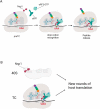
Model for Nsp1 activity in translation. (A) Translation termination in the presence of Nsp1. Nsp1 facilitates eRF1binding to the stop codon. After GTP hydrolysis and peptide release Nsp1 remains bound with the postTC. (B) Suppression of translation by Nsp1. Nsp1 binds to the 40S ribosomal subunits, which suppresses initiation of host translation. Additionally Nsp1 stimulates translation termination on the currently translating host mRNAs and displaces the 40S ribosomal subunits from the pool of the actively translating ribosomes. This prevents new rounds of host translation. Created with BioRender.com

References

    1. Zhu N, Zhang D, Wang W, et al. A novel coronavirus from patients with pneumonia in China, 2019. N Engl J Med. 2020;382:727–733. - PMC - PubMed
    1. Kim D, Lee JY, Yang JS, et al. The architecture of SARS-CoV-2 transcriptome. Cell. 2020b;181:914–921.e10. - PMC - PubMed
    1. Wu A, Peng Y, Huang B, et al. Genome composition and divergence of the novel coronavirus (2019-nCoV) originating in China. Cell Host Microbe. 2020;27:325–328. - PMC - PubMed
    1. Zhou P, Lou YX, Wang XG, et al. A pneumonia outbreak associated with a new coronavirus of probable bat origin. Nature. 2020;579:270–273. - PMC - PubMed
    1. Yuan S, Peng L, and Park JJ, et al. Nonstructural protein 1 of SARS-CoV-2 is a potent pathogenicity factor redirecting host protein synthesis machinery toward viral RNA. Mol Cell. 2020;80:1055–1066.e6 . - PMC - PubMed

Publication types

MeSH terms