. 2021 Dec 1;71(6):512-520.
doi: 10.30802/AALAS-CM-21-000061. Epub 2021 Nov 18.

Degenerative Osteoarthropathy in Laboratory Housed Xenopus (Silurana) tropicalis

Affiliations

Degenerative Osteoarthropathy in Laboratory Housed Xenopus (Silurana) tropicalis

Mingyun Zhang et al. Comp Med. .

Abstract

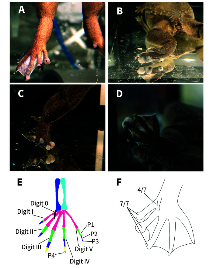
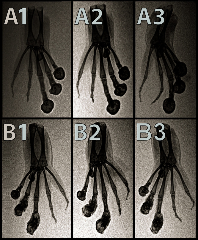
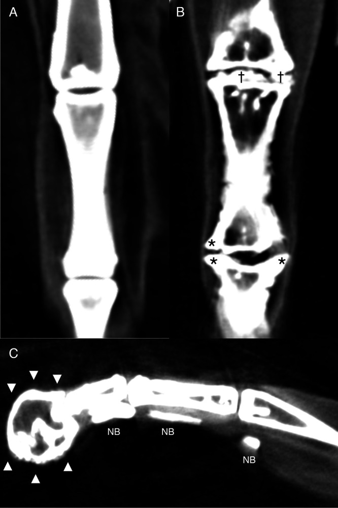
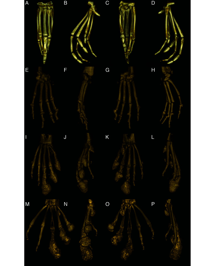
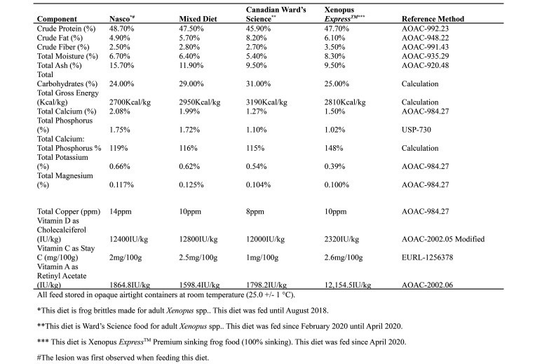
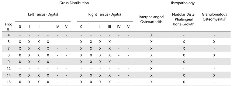
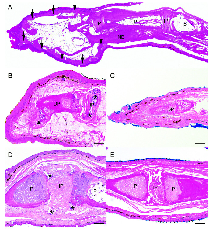
In this case study, 15 adult laboratory Xenopus (Silurana) tropicalis (7 adult males and 8 adult females) were examined for nodular enlargements of the clawed digits (digits 0, I, II, and III) on the hind feet. Radiographs showed smoothly margined, rounded, peripherally mineralized lesions arising from the distal phalanges of digits 0-III with osteoproductive and osteolytic components in all frogs. Micro computed tomography (microCT) scans further revealed interphalangeal (IP), metacarpophalangeal (MCP), and metatarsophalangeal (MTP) joint osteoarthritis characterized by periarticular new bone formation, rounded mineral foci both peripherally and centrally within the joints, and more rarely, linear mineralization palmar/plantar to the joints in the flexor tendons. In the nonclawed digits, the shape of the distal phalanx was variably distorted and both subluxation and malangulation of IP joints were identified. Histologically, nodules corresponded to a peripheral rim of mature cortical bone surrounding central adipose tissue, scattered hematopoietic elements, and residual bone of the distal phalanx. Occasionally, the peripheral rim of cortical bone extended proximally to encompass the distal aspect of adjacent phalanx. MCP, MTP and IP joint spaces of most digits exhibited widespread osteoarthritis characterized by periarticular cartilaginous or osseous metaplasia, bony remodeling, and less frequently, granulomatous osteomyelitis. Nutritional analyses of the feed did not indicate imbalances nor were the lesions consistent with metabolic bone disease. The exact etiopathogenesis of these lesions is unknown; however, we hypothesize that the osteoarthritic changes are due to a combination of the frogs' mature age, the unique structure of the Xenopus spp. claw, genetics and biomechanical forces on the digits and distal phalanges of the hind feet.


Figures

Figure 1.
Figure 1.
The gross figures of the feet of affected frogs. Four X. tropicalis displaying nodules on the distal aspects of the digits of the hindlimbs (A-D). The bone anatomy of a normal hindlimb (E). The normal X. tropicalis hind foot is characterized by: digit 0 shown in black, metatarsals in pink, tibiae in sky blue, fibulae in blue, first phalanx (P1) in green, second phalanx (P2) in purple, third phalanx (P3) in yellow, fourth phalanx (P4) in royal blue (on digit IV). Claws are only present on digits 0, I, II, and III. Note the normal, triangular-shaped distal phalanx. Hind foot diagram demonstrating which digits are the most affected (F). Seven frogs submitted directed to Stanford Necropsy Laboratory displayed nodules on the distal end of digits I, II, III of the hindlimbs. None of the frogs displayed nodules on digits IV or V.
Figure 2.
Figure 2.
Dorsoventral (A1), ventrodorsal oblique (A2), and ventrodorsal (A3) inverted-grayscale radiographs of the left hindfoot. Dorsoventral (B1), ventrodorsal oblique (B2), ventrodorsal (B3) inverted-grayscale radiographs of the right hind foot of an affected X. tropicalis. Smoothly margined, rounded peripherally mineralized lesions are present on the distal aspects of digits 0, I, II, and III. Note the malangulation of P4 and P3 on digit IV. Note the malangulation of P3 and P2 on digit V.
Figure 3.
Figure 3.
Reconstructed dorsal plane CT image of a normal MTP and IP joint (A), reconstructed dorsal plane CT image of a severely affected MTP joint and IP joint (B) with periarticular new bone formation (asterisks *) and new bone formation within the joint space (daggers †), and reconstructed sagittal plane CT image of a severely affected digit (C) with a misshaped distal phalanx surrounded by a rim of new bone (arrowheads xxx) and linear mineralization plantar to the digit within the extensor tendons (NB).
Figure 4.
Figure 4.
Reconstructed 3D volume renderings of a normal pes (A-D), a severely affected manus (E-H), a severely affected pes (I-L), and the most severely affected pes (M-P). A, E, I and M are dorsal views; B, F, J, and N are medial views, C, G, K, and O are palmar/plantar views, and D, H, L, and P are lateral views. A-D: Normal pes. E-H: Severely affected manus with severe periarticular and articular new bone formation in multiple MCP and IP joints with malangulation and subluxation of the IP joint. I-L: Severely affected pes that has rounded peripherally mineralized lesions on digits 0-III which extend proximal to the IP. There is severe linear mineralization plantar to digits I-IV in the flexor tendons. Similar to the manus, there is periarticular and articular new bone formation in the MTP and IP joints in the nonclawed digits and of the clawed digits proximal to the rounded lesions. M-P: Most severely affected pes with rounded peripherally mineralized lesions extending to the proximal phalanx, linear mineralization plantar to digits I-III, and severe periarticular new bone formation.
Figure 5.
Figure 5.
Nutritional analysis of Nasco adult frog brittle, mix of Nasco adult frog brittle and Ward’s Science frog brittle, Ward’s Science frog brittle, and Xenopus Express Xenopus diet content. Analysis was performed by Cornerstone laboratories, LLC (Cornerstone Laboratories, LLC, Memphis, TN).
Figure 6.
Figure 6.
Gross distribution and histopathologic characteristics of phalangeal nodules in Xenopus tropicalis.
Figure 7.
Figure 7.
Histologic lesions in tarsal digits 0, I, II, and III of Xenopus tropicalis. Sagittal section of affected tarsal digit III (A). Nodular lesions comprised a peripheral rim of cortical bone (black arrows ↓) that occasionally extended proximally to form a layer of new bone (NB) surrounding preexisting bone of the adjacent phalanx (P). Centrally, the nodule was composed of mature adipose tissue and scattered hematopoietic elements. H and E, scale bar = 1 mm. Bony remodeling of the distal phalanx and IP osteoarthritis (B). The distal phalanx (DP) is irregularly contoured with fibrocartilaginous metaplasia extending from the distal aspect (arrowhead xxx). Myxedematous stroma surrounds the DP. The IP joint space contains abundant mineral (asterisks *). Note: The adjacent phalanx is out of the plane of section. H and E, scale bar = 100 µm. Unaffected DP (C). H and E, scale bar = 100 µm. An affected IP joint space exhibiting incongruity of adjacent phalanges (P) with IP mineralization (asterisks *) (D). H and E, scale bar = 100 µm. Normal adjacent phalanges, IP joint space (E). H and E, scale bar = 100 µm.

References

    1. Bissegger S, Langlois VS. 2016. Androgens modulate gene expression and specific DNA methylation pattern of steroid 5□-reductases in the frog Silurana tropicalis. Gen Comp Endocrinol 234:123–132. 10.1016/j.ygcen.2016.03.021. - DOI - PubMed
    1. Campbell DE, Langlois VS. 2018. Expression of sf1 and dax-1 are regulated by thyroid hormones and androgens during Silurana tropicalis early development. Gen Comp Endocrinol 259:34–44. 10.1016/j.ygcen.2017.10.017. - DOI - PubMed
    1. Chu C-W, Masak G, Yang J, Davidson LA. 2020. From biomechanics to mechanobiology: Xenopus provides direct access to the physical principles that shape the embryo. Curr Opin Genet Dev 63:71–77. 10.1016/j.gde.2020.05.011. - DOI - PMC - PubMed
    1. de Berker D. 2013. Nail anatomy. Clin Dermatol 31:509–515. 10.1016/j.clindermatol.2013.06.006. - DOI - PubMed
    1. Dumont JN. 1972. Oogenesis in Xenopus laevis (Daudin). I. Stages of oocyte development in laboratory maintained animals. J Morphol 136:153–179. 10.1002/jmor.1051360203. - DOI - PubMed

Publication types