. 2021 Nov 18;12(1):6668.
doi: 10.1038/s41467-021-27062-5.

MDA5 disease variant M854K prevents ATP-dependent structural discrimination of viral and cellular RNA

Affiliations

MDA5 disease variant M854K prevents ATP-dependent structural discrimination of viral and cellular RNA

Qin Yu et al. Nat Commun. .

Abstract

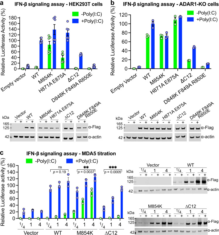
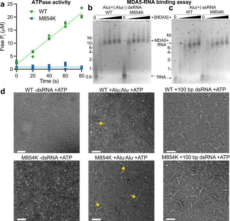
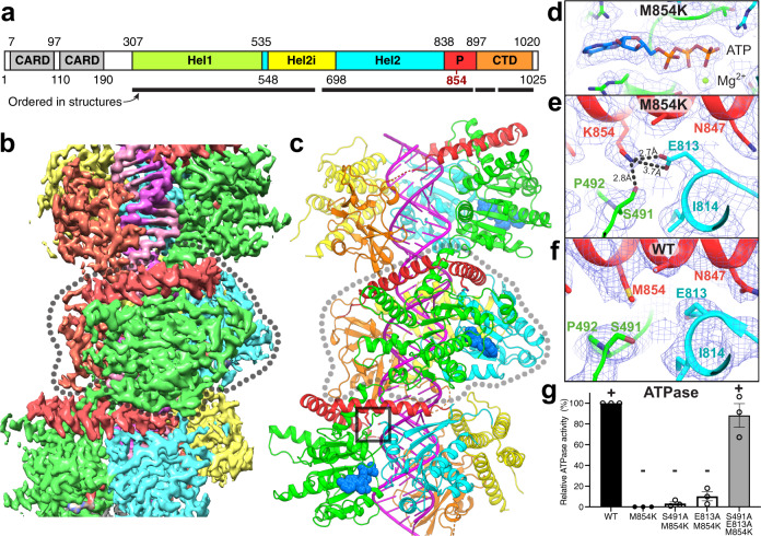
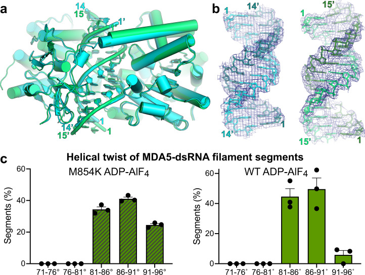
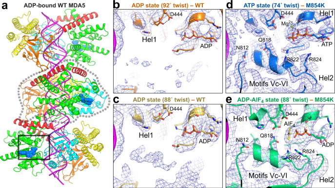
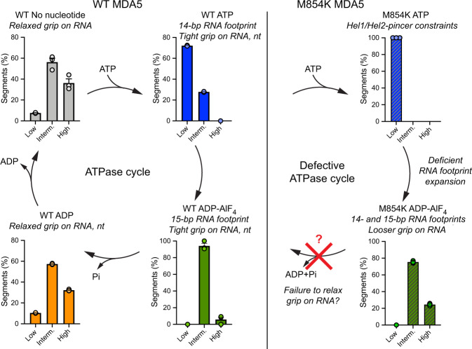
Our innate immune responses to viral RNA are vital defenses. Long cytosolic double-stranded RNA (dsRNA) is recognized by MDA5. The ATPase activity of MDA5 contributes to its dsRNA binding selectivity. Mutations that reduce RNA selectivity can cause autoinflammatory disease. Here, we show how the disease-associated MDA5 variant M854K perturbs MDA5-dsRNA recognition. M854K MDA5 constitutively activates interferon signaling in the absence of exogenous RNA. M854K MDA5 lacks ATPase activity and binds more stably to synthetic Alu:Alu dsRNA. CryoEM structures of MDA5-dsRNA filaments at different stages of ATP hydrolysis show that the K854 sidechain forms polar bonds that constrain the conformation of MDA5 subdomains, disrupting key steps in the ATPase cycle- RNA footprint expansion and helical twist modulation. The M854K mutation inhibits ATP-dependent RNA proofreading via an allosteric mechanism, allowing MDA5 to form signaling complexes on endogenous RNAs. This work provides insights on how MDA5 recognizes dsRNA in health and disease.


Conflict of interest statement

Y.M. is a consultant for Related Sciences LLC and has profits interests in Danger Bio LLC. The remaining authors declare no competing interests.

Figures

Fig. 1
Fig. 1. MDA5 M854K is constitutively active in a cell-based IFN-β reporter assay.
a IFN-β reporter signaling assay in HEK cells. Luciferase activity is normalized against WT MDA5 + Poly(I:C) as 100%. n = 6 distinct samples. Lower panel: Western blots showing the expression level of the human MDA5 mutants in HEK cells. The FLAG tag on each mutant was detected with an anti-FLAG antibody. “−”, without Poly(I:C) induction; “+”, with Poly(I:C) induction. b IFN-β reporter assay in ADAR1-KO HEK cells. Lower panel: Western blots showing the expression level of the human MDA5 mutants in ADAR1-KO cells. n = 3 distinct samples. c IFN-β reporter assay with titration of protein expression level in HEK cells. ¼, 1, and 4 indicate the fold of plasmids transfected. ns, P > 0.05; **,P < 0.01; ***P < 10−3 (n = 3 distinct samples, ordinary one-way ANOVA). WT MDA5 shows a switch-like “On-Off” activation mode for signaling. The mutants show signal activation proportional to their expression level, at low dsRNA abundance without poly(I:C). Lower panel: Western blots showing the expression level of the human MDA5 mutants in titration experiments. All error bars in this figure represent SEM between measurements, centered on the mean. Source data are provided as a Source Data file. All cell-based assays were repeated independently at least three times with similar results.
Fig. 2
Fig. 2. The ATPase, RNA binding, and filament-forming activities of MDA5 M854K.
a ATPase assay of WT MDA5 and M854K MDA5 with 1 kb dsRNA. The assay was performed in triplicate with three independently purified batches of MDA5 protein. b Gel-shift assay (EMSA) of WT or M854K MDA5 (0, 0.2, 0.4, 0.6, 0.8, 1 µM) mixed with 5 ng/μl annealed Alu(+):Alu(−) dsRNA, followed by addition of 6 mM ATP. Agarose gels were stained for RNA with SYBR Gold. The M854K variant binds to Alu(+):Alu(−) with higher affinity than WT MDA5 with ATP present. c EMSA of increasing concentrations of WT or M854K MDA5 after incubation with 5 ng/μl Alu(+) ssRNA performed under the same conditions as in (b). The M854K variant binds ssRNA with a similar affinity as WT MDA5 with ATP present. d Negative-stain electron micrographs of M854K and WT MDA5 with ATP and Alu(+):Alu(−) dsRNA, 100-bp dsRNA, or without dsRNA. Arrows indicate MDA5-Alu:Alu complexes. Scale bar, 100 nm. Micrographs are representative of at least six images collected for each condition (see Supplementary Fig. 2 for additional micrographs). ATPase assays and EMSAs were repeated independently at least three times with similar results. Source data are provided as a Source Data file.
Fig. 3
Fig. 3. CryoEM structure of the ATP-bound M854K MDA5-dsRNA filament.
a Domain architecture of human/mouse MDA5. CARD caspase recruitment domain; Hel1 and Hel2 first and second RecA-like helicase domains; Hel2i, Hel2 insert domain; P pincer domain; CTD C-terminal domain. The same color code is used in panels (be), Fig. 5a and Supplementary Fig. 4. b 3D density map of the ATP-bound M854K MDA5-dsRNA filament at 2.8 Å overall resolution, colored by domain as in (a), with RNA in magenta. An MDA5 protomer is outlined in dashed gray. c Overview of the atomic model of the ATP-bound M854K MDA5-dsRNA filament. The central protomer of three MDA5 protomers is outlined in light gray. ATP is shown in sphere representation. The region surrounding residue 854 is boxed. d, e Closeups of cryoEM density maps: the bound ATP, d, and the region surrounding residue 854, showing polar contacts formed by Lys854 with residues from the Hel1 and Hel2 domains, (e). Polar contacts formed by Lys854 are shown as dashed lines. f Closeup of the region surrounding residue 854 in the previously reported structure of the WT MDA5-dsRNA filament with AMPPNP bound. g Relative ATPase activities of S491A/M854K, E813A/M854K, and S491A/E813A/M854K mutants, normalized to WT (100%) and M854K (0%). Error bars represent SEM between measurements, centered on the mean. (n = 3 independent samples; t = 15 min). ATPase assays were repeated in independent experiments three times with similar results. Source data are provided as a Source Data file.
Fig. 4
Fig. 4. CryoEM structures of ADP-AlF4-bound M854K MDA5-dsRNA filaments.
a Superposition of two atomic models of ADP-AlF4-bound M854K MDA5-dsRNA filaments representing the most populated classes of 3D cryoEM image reconstructions. The class representing the majority of filament segments (Class 167k, cyan) contains 14 bp RNA in the asymmetric unit; the minority structural class (Class 62k, green) contains 15 bp RNA. b Atomic models and 3D density maps for the dsRNA for the two structural classes of ADP-AlF4-bound M854K MDA5-dsRNA filaments. Left, Class 167k, 6.8-σ contour level in PyMol; Right, Class 62k, 6.0-σ contour level. c Histograms showing the distributions of filament segments as a function of helical twist for M854K and WT MDA5-dsRNA with ADP-AlF4 bound. The twist distribution for WT is shown for reference and was reported previously. The distributions are from 3D classifications performed with ten classes per dataset. Error bars represent the SEM between 3D classification calculations, centered on the mean (n = 3 independent calculations). Source data are provided as a Source Data file.
Fig. 5
Fig. 5. CryoEM structures of WT MDA5-dsRNA filaments in complex with ADP.
a Atomic model of the WT MDA5-dsRNA filament with ADP bound and 92˚ helical twist, determined from a cryoEM map at 3.35 Å overall resolution. The central protomer of three MDA5 protomers is outlined in dashed gray. The region around the ATP binding site and motifs Vc-VI, shown in (b), is boxed. The color code is the same as in Fig. 1. b, c CryoEM density at the Hel1-Hel2 interface in ADP-bound WT MDA5-dsRNA filaments. b High-helical twist (92˚) structure; c intermediate-twist (88˚) structure. d, e CryoEM density at the Hel1-Hel2 interface in ATP-bound M854K MDA5-dsRNA filaments with an Mg2+ ion shown as a green sphere, d, and ADP-AlF4-bound M854K MDA5-dsRNA filaments (Class 62k), e Motifs Vc-VI become disordered in the transition from the ADP-AlF4 state to the ADP state. The same contour level of 5.5 σ in PyMol was used for panels be.
Fig. 6
Fig. 6. Helical twist of MDA5 filaments in the ATPase cycle and proposed effect of M854K on the twist.
Histograms showing the distributions of 3D cryoEM reconstructions as a function of helical twist for WT and M854K MDA5-dsRNA filaments with ATP, ADP-AlF4, ADP, or no nucleotide bound. The distributions shown are from 3D classifications performed with ten classes per dataset. Error bars represent SEM between 3D classification calculations, centered on the mean (n = 3 independent calculations). The histograms for nucleotide-free, ATP-bound, and ADP-AlF4-bound WT MDA5-RNA filaments are from data reported previously. Low, twist = 71˚–81˚; Interm., twist = 81˚–91˚; High, twist = 91˚–96˚. “nt”, ANP nucleotide. See also Supplementary Fig. 8. Source data are provided as a Source Data file.

References

    1. Kato H, et al. Differential roles of MDA5 and RIG-I helicases in the recognition of RNA viruses. Nature. 2006;441:101–105. doi: 10.1038/nature04734. - DOI - PubMed
    1. Kato H, et al. Length-dependent recognition of double-stranded ribonucleic acids by retinoic acid-inducible gene-I and melanoma differentiation-associated gene 5. J. Exp. Med. 2008;205:1601–1610. doi: 10.1084/jem.20080091. - DOI - PMC - PubMed
    1. Goubau D, et al. Antiviral immunity via RIG-I-mediated recognition of RNA bearing 5’-diphosphates. Nature. 2014;514:372–375. doi: 10.1038/nature13590. - DOI - PMC - PubMed
    1. Peisley A, et al. Cooperative assembly and dynamic disassembly of MDA5 filaments for viral dsRNA recognition. Proc. Natl Acad. Sci. USA. 2011;108:21010–21015. doi: 10.1073/pnas.1113651108. - DOI - PMC - PubMed
    1. Berke IC, Modis Y. MDA5 cooperatively forms dimers and ATP-sensitive filaments upon binding double-stranded RNA. EMBO J. 2012;31:1714–1726. doi: 10.1038/emboj.2012.19. - DOI - PMC - PubMed

Publication types

MeSH terms