

Developing a pro-angiogenic placenta derived amniochorionic scaffold with two exposed basement membranes as substrates for cultivating endothelial cells
- PMID: 34795361
- PMCID: PMC8602627
- DOI: 10.1038/s41598-021-01922-y
Developing a pro-angiogenic placenta derived amniochorionic scaffold with two exposed basement membranes as substrates for cultivating endothelial cells
Abstract
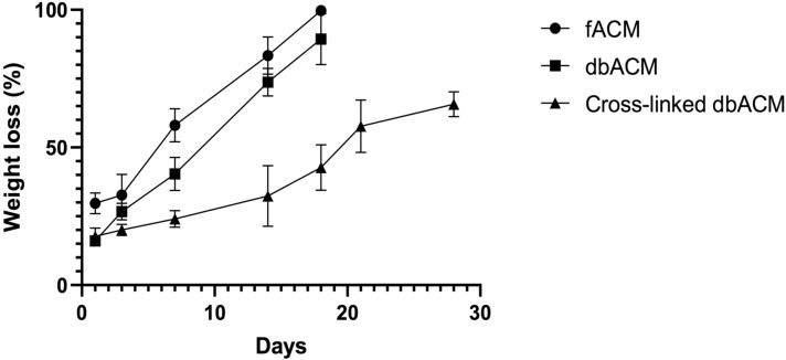
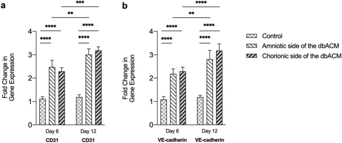
Decellularized and de-epithelialized placenta membranes have widely been used as scaffolds and grafts in tissue engineering and regenerative medicine. Exceptional pro-angiogenic and biomechanical properties and low immunogenicity have made the amniochorionic membrane a unique substrate which provides an enriched niche for cellular growth. Herein, an optimized combination of enzymatic solutions (based on streptokinase) with mechanical scrapping is used to remove the amniotic epithelium and chorion trophoblastic layer, which resulted in exposing the basement membranes of both sides without their separation and subsequent damages to the in-between spongy layer. Biomechanical and biodegradability properties, endothelial proliferation capacity, and in vivo pro-angiogenic capabilities of the substrate were also evaluated. Histological staining, immunohistochemistry (IHC) staining for collagen IV, and scanning electron microscope demonstrated that the underlying amniotic and chorionic basement membranes remained intact while the epithelial and trophoblastic layers were entirely removed without considerable damage to basement membranes. The biomechanical evaluation showed that the scaffold is suturable. Proliferation assay, real-time polymerase chain reaction for endothelial adhesion molecules, and IHC demonstrated that both side basement membranes could support the growth of endothelial cells without altering endothelial characteristics. The dorsal skinfold chamber animal model indicated that both side basement membranes could promote angiogenesis. This bi-sided substrate with two exposed surfaces for cultivating various cells would have potential applications in the skin, cardiac, vascularized composite allografts, and microvascular tissue engineering.
© 2021. The Author(s).
Conflict of interest statement
The authors declare no competing interests.
Figures


References
-
- Deus IA, Mano JF, Custódio CA. Perinatal tissues and cells in tissue engineering and regenerative medicine. Acta Biomater. 2020;110:1–14. - PubMed
-
- Swim MM, Albertario A, Iacobazzi D, Caputo M, Ghorbel MT. Amnion-based scaffold with enhanced strength and biocompatibility for in vivo vascular repair. Tissue Eng. Part A. 2019;25:603–619. - PubMed
-
- Roy A, Griffiths S. Intermediate layer contribution in placental membrane allografts. J. Tissue Eng. Regen. Med. 2020;14:1126–1135. - PubMed
-
- Koob TJ, Lim JJ, Zabek N, Massee M. Cytokines in single layer amnion allografts compared to multilayer amnion/chorion allografts for wound healing. J. Biomed. Mater. Res. B Appl. Biomater. 2015;103:1133–1140. - PubMed
Publication types
MeSH terms
Substances
Grants and funding
LinkOut - more resources
Full Text Sources
Miscellaneous
