Clinical Trial
. 2022 Mar;126(4):598-605.
doi: 10.1038/s41416-021-01623-3. Epub 2021 Nov 18.

Characterising 18F-fluciclovine uptake in breast cancer through the use of dynamic PET/CT imaging

Affiliations
Clinical Trial

Characterising 18F-fluciclovine uptake in breast cancer through the use of dynamic PET/CT imaging

N P Scott et al. Br J Cancer. 2022 Mar.

Abstract

Background: 18F-fluciclovine is a synthetic amino acid positron emission tomography (PET) radiotracer that is approved for use in prostate cancer. In this clinical study, we characterised the kinetic model best describing the uptake of 18F-fluciclovine in breast cancer and assessed differences in tracer kinetics and static parameters for different breast cancer receptor subtypes and tumour grades.

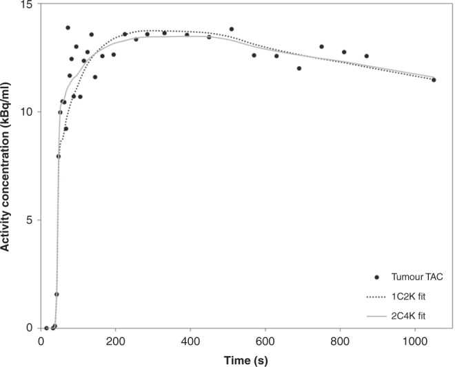
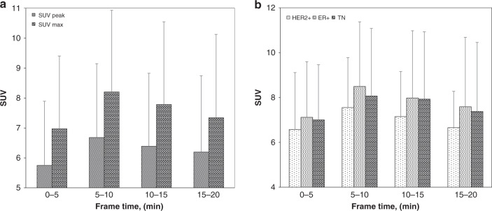
Methods: Thirty-nine patients with pathologically proven breast cancer underwent 20-min dynamic PET/computed tomography imaging following the administration of 18F-fluciclovine. Uptake into primary breast tumours was evaluated using one- and two-tissue reversible compartmental kinetic models and static parameters.

Results: A reversible one-tissue compartment model was shown to best describe tracer uptake in breast cancer. No significant differences were seen in kinetic or static parameters for different tumour receptor subtypes or grades. Kinetic and static parameters showed a good correlation.

Conclusions: 18F-fluciclovine has potential in the imaging of primary breast cancer, but kinetic analysis may not have additional value over static measures of tracer uptake.

Clinical trial registration: NCT03036943.


Conflict of interest statement

At the time of submission, EJT is an employee of Blue Earth Diagnostics. The views expressed in this publication are those of the authors and not necessarily those of the NHS, the NIHR, HEE or the Department of Health and Social Care. The authors declare no competing interests.

Figures

Fig. 1
Fig. 1. Summed 5- to 10-min dynamic 18F-fluciclovine PET and fused PET/CT images of a patient with oestrogen receptor-positive IDC.
The primary tumour is indicated by the arrows in the axial plane of the fused PET/CT images.
Fig. 2
Fig. 2. Graphical representation of the time course of tumour tracer uptake in an example patient in the study.
One- and two-tissue compartment models have been fitted to the tracer uptake in the tumour.
Fig. 3
Fig. 3. SUVs of 18F-fluciclovine in breast tumours at varying time points.
a SUVmax and SUVpeak uptake of 18F-fluciclovine, b SUVmax uptake broken down by receptor subtype. Error bars represent the standard deviation from the mean value.
Fig. 4
Fig. 4. Correlations of SUVmax against kinetic parameters for 18F-fluciclovine in breast tumours.
a K1 and b  volume of distribution (K1/k2) plotted against SUVmax. Goodness-of-fit lines are displayed alongside the corresponding goodness of fit value, R.
Fig. 5
Fig. 5. 18F-fluciclovine uptake across tumour receptor subtypes.
a SUVmax, b SUVpeak, c K1 and d volume of distribution plotted for oestrogen receptor positive only (ER+), human epidermal growth factor receptor positive (HER2+) and triple-negative (TN) hormone receptor subtypes.
Fig. 6
Fig. 6. 18F-fluciclovine uptake in patients taking metformin.
Mean values of a SUVmax, b SUVpeakc K1 and d volume of distribution plotted for patients who were on and off metformin. Error bars represent the standard deviation from the mean.

References

    1. Hyo SL, Yoon W, Tae WC, Jae KK, Jin GP, Heoung KK. FDG PET/CT for the detection and evaluation of breast diseases: usefulness and limitations. Radiographics. 2007;27:S197–213. doi: 10.1148/rg.27si075507. - DOI - PubMed
    1. Wahl RL, Siegel BA, Coleman RE, Gatsonis CG. Prospective multicenter study of axillary nodal staging by positron emission tomography in breast cancer: a report of the staging breast cancer with PET study group. J Clin Oncol. 2004;22:277–85. doi: 10.1200/JCO.2004.04.148. - DOI - PubMed
    1. Aroldi F, Lord SR. Window of opportunity clinical trial designs to study cancer metabolism. Br J Cancer. 2020;122:45–51. doi: 10.1038/s41416-019-0621-4. - DOI - PMC - PubMed
    1. Plathow C, Weber WA. Tumor cell metabolism imaging. Journal of nuclear medicine: official publication. Soc Nucl Med. 2008;49:43S–63S. doi: 10.2967/jnumed.107.045930. - DOI - PubMed
    1. Mcconathy J. 18F-Fluciclovine (FACBC) and its potential use for breast cancer imaging. J Nucl Med. 2016;57:1329–30. - PubMed

Publication types

MeSH terms

Associated data