

Transcription Factor Control of Lymphatic Quiescence and Maturation of Lymphatic Neovessels in Development and Physiology
- PMID: 34795596
- PMCID: PMC8593113
- DOI: 10.3389/fphys.2021.672987
Transcription Factor Control of Lymphatic Quiescence and Maturation of Lymphatic Neovessels in Development and Physiology
Abstract
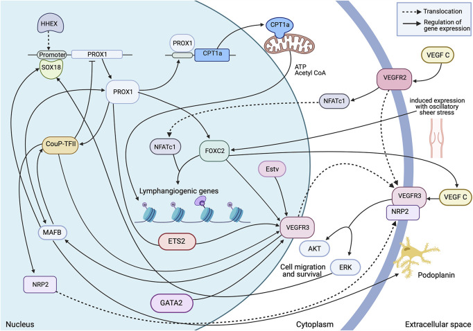
The lymphatic system is a vascular system comprising modified lymphatic endothelial cells, lymph nodes and other lymphoid organs. The system has diverse, but critical functions in both physiology and pathology, and forms an interface between the blood vascular and immune system. It is increasingly evident that remodelling of the lymphatic system occurs alongside remodelling of the blood microvascular system, which is now considered a hallmark of most pathological conditions as well as being critical for normal development. Much attention has focussed on how the blood endothelium undergoes phenotypic switching in development and disease, resulting in over two decades of research to probe the mechanisms underlying the resulting heterogeneity. The lymphatic system has received less attention, and consequently there are fewer descriptions of functional and molecular heterogeneity, but differential transcription factor activity is likely an important control mechanism. Here we introduce and discuss significant transcription factors of relevance to coordinating cellular responses during lymphatic remodelling as the lymphatic endothelium dynamically changes from quiescence to actively remodelling.
Keywords: inflammation; lymphangiogenesis; lymphatic; quiescence; transcription factor.
Copyright © 2021 Tabrizi, Ahmed, Horder, Storr and Benest.
Conflict of interest statement
The authors declare that the research was conducted in the absence of any commercial or financial relationships that could be construed as a potential conflict of interest.
Figures


Similar articles
-
Lymphangiogenesis in development and disease.Novartis Found Symp. 2007;283:87-98; discussion 98-105, 238-41. doi: 10.1002/9780470319413.ch8. Novartis Found Symp. 2007. PMID: 18300416
-
Interaction of tumor cells and lymphatic vessels in cancer progression.Oncogene. 2012 Oct 18;31(42):4499-508. doi: 10.1038/onc.2011.602. Epub 2011 Dec 19. Oncogene. 2012. PMID: 22179834 Review.
-
Neutrophil Interactions with the Lymphatic System.Cells. 2021 Aug 17;10(8):2106. doi: 10.3390/cells10082106. Cells. 2021. PMID: 34440875 Free PMC article. Review.
-
Molecular mechanisms of lymphatic metastasis in solid tumors of the gastrointestinal tract.Int J Clin Exp Pathol. 2012;5(7):614-23. Epub 2012 Sep 5. Int J Clin Exp Pathol. 2012. PMID: 22977656 Free PMC article. Review.
-
Organ-specific lymphatic vasculature: From development to pathophysiology.J Exp Med. 2018 Jan 2;215(1):35-49. doi: 10.1084/jem.20171868. Epub 2017 Dec 14. J Exp Med. 2018. PMID: 29242199 Free PMC article. Review.
Cited by
-
Molecular and metabolic orchestration of the lymphatic vasculature in physiology and pathology.Nat Commun. 2023 Dec 16;14(1):8389. doi: 10.1038/s41467-023-44133-x. Nat Commun. 2023. PMID: 38104163 Free PMC article. Review.
-
Dental pulp lymphatic vessel dynamics during tooth development and pulp stimulation in rodents.Int Endod J. 2025 Aug;58(8):1197-1210. doi: 10.1111/iej.14244. Epub 2025 Apr 25. Int Endod J. 2025. PMID: 40277146 Free PMC article.
-
Inflammatory Alterations to Renal Lymphatic Endothelial Cell Gene Expression in Mouse Models of Hypertension.Kidney Blood Press Res. 2024;49(1):588-604. doi: 10.1159/000539721. Epub 2024 Jul 22. Kidney Blood Press Res. 2024. PMID: 38972305 Free PMC article.
-
Mechanisms of Myocardial Edema Development in CVD Pathophysiology.Biomedicines. 2024 Feb 19;12(2):465. doi: 10.3390/biomedicines12020465. Biomedicines. 2024. PMID: 38398066 Free PMC article. Review.
-
Robust Differentiation of Human Pluripotent Stem Cells into Lymphatic Endothelial Cells Using Transcription Factors.Cells Tissues Organs. 2024;213(6):464-474. doi: 10.1159/000539699. Epub 2024 Aug 28. Cells Tissues Organs. 2024. PMID: 39197437 Free PMC article.
References
Publication types
LinkOut - more resources
Full Text Sources