A rare case of pediatric primary central nervous system differentiating neuroblastoma: an unusual and rare intracranial primitive neuroectodermal tumor (a case report)
- PMID: 34795814
- PMCID: PMC8571934
- DOI: 10.11604/pamj.2021.40.33.30587
A rare case of pediatric primary central nervous system differentiating neuroblastoma: an unusual and rare intracranial primitive neuroectodermal tumor (a case report)
Abstract
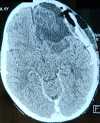
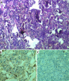
Neuroblastoma represents the most common solid extracranial tumor in children under 5, accounting for 8% to 10% of all childhood cancers. Primary central nervous system (CNS) neuroblastomas are a very rare location and only few cases are available in the literature. It was first described in 1973 by Hart and Earl as supratentorial primitive neuroectodermal tumors. Clinical presentation is highly variable and depends on the initial location of the tumor. Regarding imaging, primary brain neuroblastoma shows no pathognomonic appearance on brain computed tomography (CT) whether or not enhanced or magnetic resonance imaging (MRI). There were no standard guidelines available for the adjuvant treatment in case of primary CNS neuroblastoma. Surgery remains the main and the first tool toward these lesions. Radiotherapy associated or not to chemotherapy is offered based on patient´s age. Here, the authors report a new pediatric case of primitive central nervous system neuroblastoma revealed by an intracranial hypertension syndrome and confirmed by both histopathological and immunohistochemistry study after a gross total surgical excision. The postoperative course was uneventful and the child had good recovery.
Keywords: Neuroblastoma; case report; chemotherapy; radiotherapy; surgery.
Copyright: Mehdi Borni et al.
Conflict of interest statement
The authors declare no competing interests.
Figures


References
-
- Schwab M, Westermann F, Hero B, Berthold F. Neuroblastoma: biology and molecular and chromosomal pathology. Lancet Oncol. 2003 Aug;4(8):472–80. - PubMed
-
- Crom DB, Wilimas JA, Green AA, Pratt CB, Jenkins 3rd JJ, Behm FG. Malignancy in the neonate. Med Pediatr Oncol. 1989;17:101–4. - PubMed
-
- Michalowski MB, Rubie H, Michon J, Montamat S, Bergeron C, Coze C, et al. Neuroblastomes localisés du nouveau-né: 52 cas traités de 1990 à 1999. Arch Péd. 2004;11(7):782–8. - PubMed
-
- Brodeur GM, Maris JM. Neuroblastoma. In: Pizzo PA, Poplack DG, editors. principles and practice of pediatric oncology. 4th edition. Philadelphia: Lippincott Williams & Wilkins; 2002. pp. 895–937.
-
- Olshan AF, Bunin GR. Epidemiology of neuroblastoma. Neuroblastoma, Elsevier, Amsterdam. 2000:33–39.
Publication types
MeSH terms
LinkOut - more resources
Full Text Sources
Medical
Molecular Biology Databases
