

Intra-operative application of ultrasonography (USG) for reduction of zygomatic arch fracture
- PMID: 34795898
- PMCID: PMC8582023
- DOI: 10.1002/ccr3.5067
Intra-operative application of ultrasonography (USG) for reduction of zygomatic arch fracture
Abstract
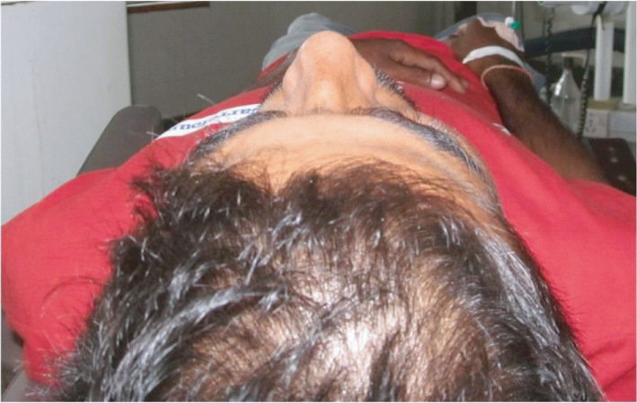
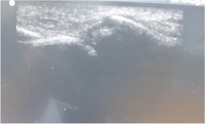
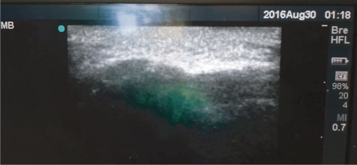
Zygomatic arch fractures are the most common facial fractures or second in frequency after the nasal fractures. The high incidence of zygomatic fractures probably relates to its prominent position in the facial skeleton; hence, it is frequently exposed to fractures. This case report presents an left-sided isolated zygomatic arch fracture after subjected to routine investigations and radiographs like submentovertex and CT scans. The patient was operated under general anesthesia for the reduction of zygomatic arch by Gille's temporal approach with the use of Ultrasound intra-operatively. Recommendation for the use of ultrasonography in the identification of zygomatic arch fractures intra-operatively operatively.
Keywords: fracture; reduction; ultrasound; zygomatic arch.
© 2021 The Authors. Clinical Case Reports published by John Wiley & Sons Ltd.
Conflict of interest statement
All the authors have no conflict of interest to declare.
Figures


References
-
- Elizabeth K, Smith SE, Frates MC, Caterson EJ. Use of high frequency ultrasound guidance for intraoperative zygomatic arch fracture reduction. J Cranio‐Max‐facSurg. 2013;24:6. - PubMed
-
- McCann J, Brocklebank M, Ayoub F. Assessment of zygomatico‐orbital complex fractures using ultrasonography. Br J OralMaxillofac Surg. 2000;38:525. - PubMed
-
- Friedrich RE, Heiland M, Bartel‐Friedrich S. Potentials of ultrasound in the diagnosis of midfacial fractures. Clin Oral Invest. 2003;7:226‐229. - PubMed
-
- Westendorff C, Gulicher D, Dammann F, Reinert S, Hoffmann J. Computer‐assisted surgical treatment of orbitozygomatic fractures. J Craniofac Surg. 2006;17:837‐842. - PubMed
-
- Pedemonte C, Saez F, Vargas I, et al. C‐arm as intraoperative control in reduction of isolated zygomatic arch fractures: a randomized clinical trial. Oral Maxillofac Surg. 2016;20:79‐83. - PubMed
Publication types
LinkOut - more resources
Full Text Sources
