Review
. 2021 Oct;13(10):6129-6140.
doi: 10.21037/jtd-2019-rts-05.

Beyond the learning curve: a review of complex cases in robotic thoracic surgery

Affiliations
Review

Beyond the learning curve: a review of complex cases in robotic thoracic surgery

Travis C Geraci et al. J Thorac Dis. 2021 Oct.

Abstract

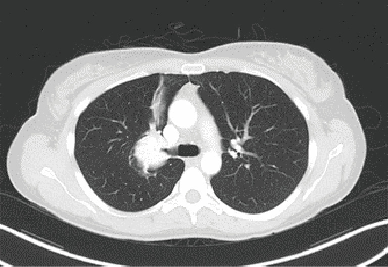
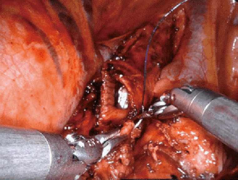
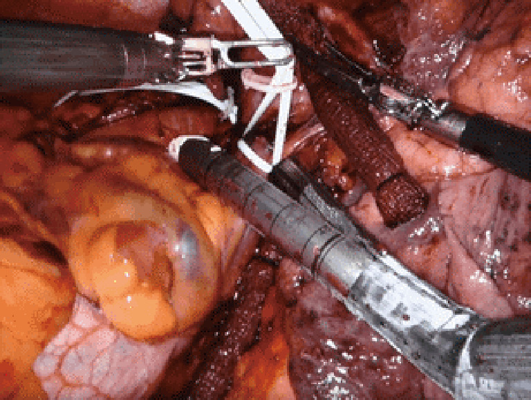
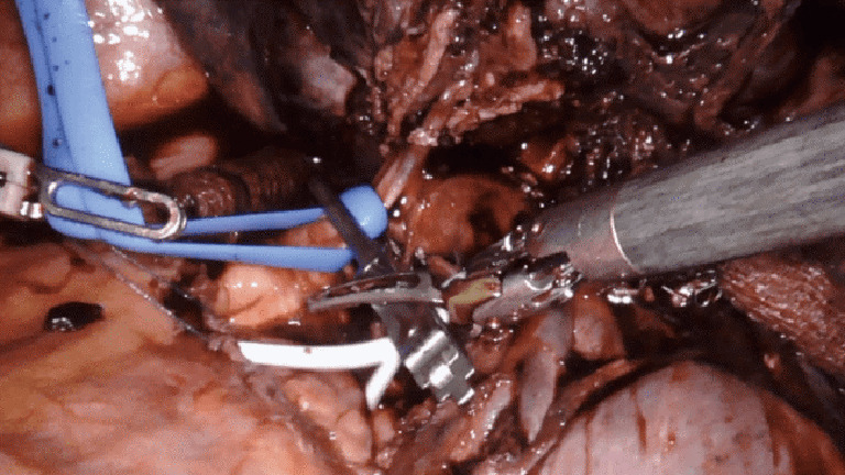
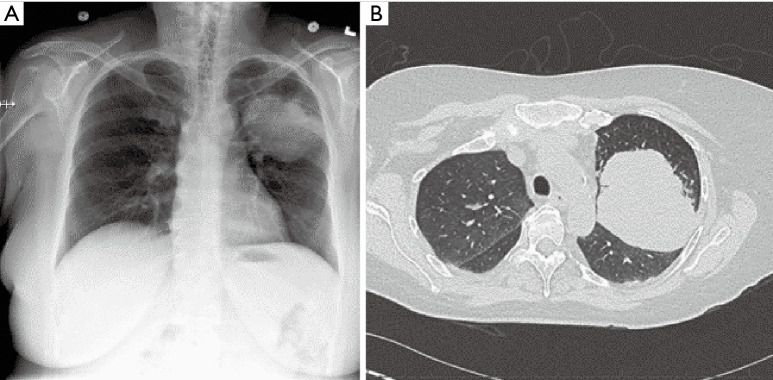
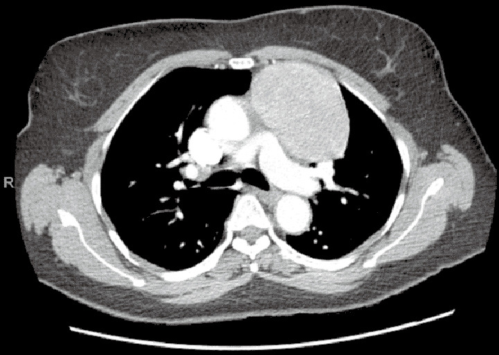
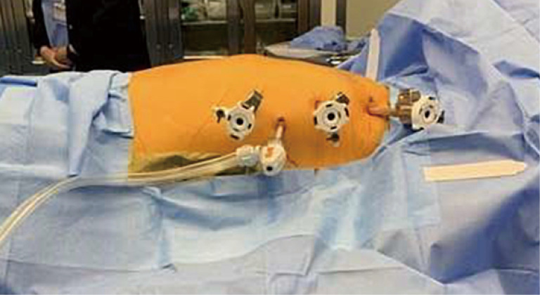
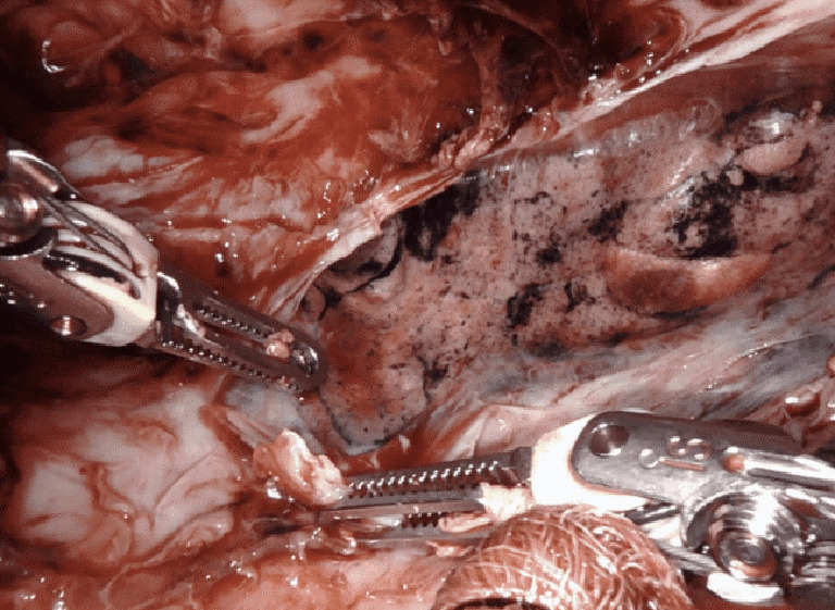
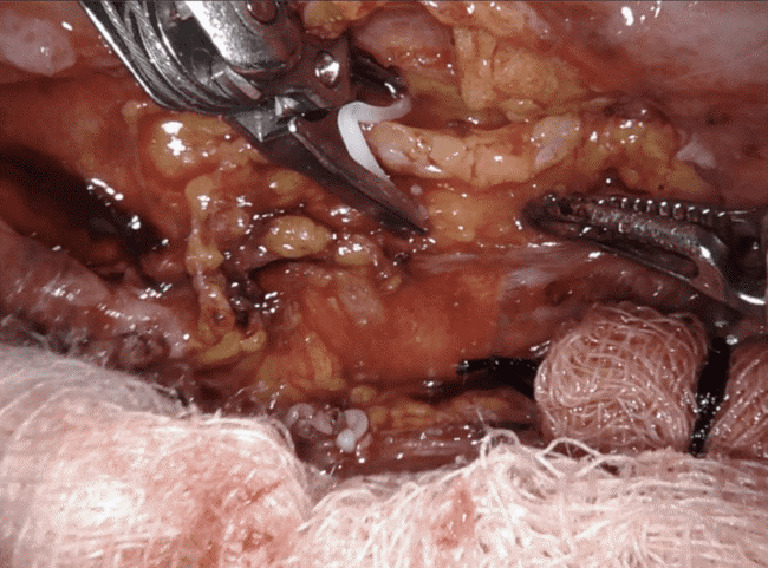
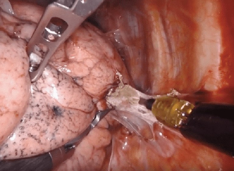
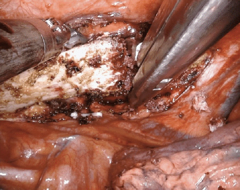
The number of thoracic surgery cases performed on the robotic platform has increased steadily over the last two decades. An increasing number of surgeons are training on the robotic system, which like any new technique or technology, has a progressive learning curve. Central to establishing a successful robotic program is the development of a dedicated thoracic robotic team that involves anesthesiologists, nurses, and bed-side assistants. With an additional surgeon console, the robot is an excellent platform for teaching. Compared to current methods of video-assisted thoracoscopic surgery (VATS), the robot offers improved wristed motion, a magnified, high definition three-dimensional vision, and greater surgeon control of the operation. These advantages are paired with integrated adjunctive technology such as infrared imaging. For pulmonary resection, these advantages of the robotic platform have translated into several clinical benefits, such as fewer overall complications, reduced pain, shorter length of stay, better postoperative pulmonary function, lower operative blood loss, and a lower 30-day mortality rate compared to open thoracotomy. With increased experience, cases of greater complexity are being performed. This review article details the process of becoming an experienced robotic thoracic surgeon and discusses a series of challenging cases in robotic thoracic surgery that a surgeon may encounter "beyond the learning curve". Nearly all thoracic surgery can now be approached robotically, including sleeve lobectomy, pneumonectomy, resection of large pulmonary and mediastinal masses, decortication, thoracic duct ligation, rib resection, and pulmonary resection after prior chest surgery and/or chemoradiation.

Keywords: Robotic surgery; learning-curve; outcomes; technique; thoracic.


Conflict of interest statement

Conflicts of Interest: All authors have completed the ICMJE uniform disclosure form (available at http://dx.doi.org/10.21037/jtd-2019-rts-05). The series “Robotic Thoracic Surgery” was commissioned by the editorial office without any funding or sponsorship. Dr. RJC reports personal fees from AstraZeneca, personal fees from Bard Davol, personal fees from Bovie Medical, personal fees from C-SATS, personal fees from ConMed, personal fees from Covidien/Medtronic, personal fees from Ethicon, personal fees from Fruit Street Health, personal fees from Google/Verb Surgical, personal fees from Intuitive Surgical, personal fees from KCI/Acelity, personal fees from Myriad Genetics, personal fees from Neomend, personal fees from Pinnacle Biologics, personal fees from ROLO-7, personal fees from Tego, personal fees from TransEnterix, outside the submitted work. Dr. CB reports personal fees from CSA Medical, outside the submitted work. Dr. MDZ reports personal fees from Intuitive Surgical, outside the submitted work. The other authors have no other conflicts of interest to declare.

Figures

Figure 1
Figure 1
CT chest, tumor obstructing the orifice of the right upper lobe bronchus. CT, computed tomography.
Figure 2
Figure 2
Bronchial anastomosis, right upper lobe sleeve lobectomy. Case: a 42-year-old woman was found to have an endobronchial carcinoid of the right upper lobe, obstructing the entire bronchus, with associated parenchymal volume loss and atelectasis. A robotic right upper lobe sleeve lobectomy with mediastinal lymphadenectomy and pleural wrap was performed.
Figure 3
Figure 3
Left intrapericardial pneumonectomy, stapling of the common pulmonary vein (background: vessel loop control of the pulmonary artery). Case: a 79-year-old man was found to have a large left hilar lung mass with compression of the left atrium and pulmonary artery. Biopsy revealed leiomyosarcoma, which was removed by left robotic pneumonectomy with intrapericardial control of the hilar vessels.
Figure 4
Figure 4
Intrapericardial control of the pulmonary artery; proximal via a bulldog clamp and vessel-loop (blue); distal control with a vessel loop (white). Case: a 54-year-old man was found to have a squamous cell lung cancer of the left upper lobe that invaded the posterior mediastinum, compressing the pulmonary artery to the left upper lobe. The patient underwent induction chemotherapy followed by robotic left upper lobectomy. Due to the tumor location compressing the pulmonary artery and extension into the mediastinum, proximal control of the pulmonary artery was obtained in the pericardium to reduce the risk of bleeding and to assure an R0 resection.
Figure 5
Figure 5
Large left upper lobe mass. (A) CXR; (B) CT scan of the chest. Case: a 53-year-old female with an 8.6-cm mass in the left upper lobe. Transthoracic biopsy revealed adenocarcinoma and an endobronchial ultrasound (EBUS) did not demonstrate cancer in the mediastinal lymph node stations 2, 4, and 7 (T4N0M0). Post neoadjuvant chemotherapy, the patient underwent robotic left upper lobectomy and superior segmentectomy of the left lower lobe (superior segment was found to be involved with tumor). CXR, chest X-ray; CT, computed tomography.
Figure 6
Figure 6
Large anterior mediastinal mass by CT scan of the chest. Case: a 65-year-old female with an 8.7-cm anterior mediastinal mass. Diagnostic evaluation revealed absence of acetylcholine receptor antibodies and normal beta-HCG and AFP levels. A robotic resection of the mediastinal mass was performed via the left chest. The pleura anterior to the mass was dissected, mobilizing the mass into the right pleural space, while moving cephalad to the level of the innominate vein. The left and right horns were dissected. Visualization and protection of the left (not involved with the mass) and right phrenic nerves was assured. Two inferior port sites were consolidated into one incision to extirpate the mass. Pathology demonstrated a type AB thymoma, 9.5 cm in diameter. CT, computed tomography; HCG, human chorionic gonadotropin; AFP, alpha-fetoprotein.
Figure 7
Figure 7
Left Port sites for resection of an anterior mediastinal mass. We utilize a 3-arm approach with an assistant port. The ports approximate the anterior axillary line with the assist port triangulated inferiorly.
Figure 8
Figure 8
Robotic decortication of the left lower lobe. Case: a 51-year-old male with a history of pneumonia, persistent pleuritic chest pain, and chronic pleural effusion was brought to the operating room for robotic decortication. The lung was mobilized with clearance of multiple pockets of loculated fluid, the lung was decorticated, and the chest was drained.
Figure 9
Figure 9
Right chest, clip application to the dissected thoracic duct. Case: a 67-year-old male post bilateral internal mammary artery bypass grafting complicated by bilateral high-output chylothorax. In the left chest, an avulsion of a major lymphatic duct was discovered and clipped. In the right chest, multiple small branches were clipped high in the anterior mediastinum adjacent the origin of the right internal mammary artery. The thoracic duct was localized posteriorly, clipped and ligated.
Figure 10
Figure 10
Robotic take-down of pleural adhesions after prior surgery and chemotherapy. Case: a 70-year-old female with a history of prior thoracotomy with left upper lobectomy followed by adjuvant chemotherapy, was found to have an enlarging left lower lobe lesion on surveillance imaging. The nodule was localized using navigational bronchoscopy with indocyanine green contrast and a non-anatomic wedge resection was performed. A limited lymphadenectomy was completed given prior lymph node dissection.
Figure 11
Figure 11
Ronguer division of the first rib from a robotic approach. Case: a 22-year-old male presented with refractory right arm swelling with paresthesias and diagnosed with right subclavian venous thrombosis. The patient underwent robotic resection of the right 1st rib with division of the scalene muscles.

References

    1. Oh DS, Reddy RM, Gorrepati ML, et al. Robotic-Assisted, Video-Assisted Thoracoscopic and Open Lobectomy: Propensity-Matched Analysis of Recent Premier Data. Ann Thorac Surg 2017;104:1733-40. 10.1016/j.athoracsur.2017.06.020 - DOI - PubMed
    1. Cerfolio RJ, Bryant AS, Minnich DJ. Starting a robotic program in general thoracic surgery: why, how, and lessons learned. Ann Thorac Surg 2011;91:1729-36; discussion 1736-7. 10.1016/j.athoracsur.2011.01.104 - DOI - PubMed
    1. Baldonado JJAR, Amaral M, Garrett J, et al. Credentialing for robotic lobectomy: what is the learning curve? A retrospective analysis of 272 consecutive cases by a single surgeon. J Robot Surg 2019;13:663-9. 10.1007/s11701-018-00902-1 - DOI - PubMed
    1. Zhang Y, Liu S, Han Y, et al. Robotic Anatomical Segmentectomy: An Analysis of the Learning Curve. Ann Thorac Surg 2019;107:1515-22. 10.1016/j.athoracsur.2018.11.041 - DOI - PubMed
    1. Feczko AF, Wang H, Nishimura K, et al. Proficiency of Robotic Lobectomy Based on Prior Surgical Technique in The Society of Thoracic Surgeons General Thoracic Database. Ann Thorac Surg 2019;108:1013-20. 10.1016/j.athoracsur.2019.04.046 - DOI - PubMed