

doi: 10.21037/jtd.2019.12.68.
Robotic pulmonary segmentectomy
Affiliations
- PMID: 34795969
- PMCID: PMC8575848
- DOI: 10.21037/jtd.2019.12.68
Item in Clipboard
Robotic pulmonary segmentectomy
J Thorac Dis.
2021 Oct.
No abstract available
Conflict of interest statement
Conflicts of Interest: All authors have completed the ICMJE uniform disclosure form (available at http://dx.doi.org/10.21037/jtd.2019.12.68). The series “Robotic Thoracic Surgery” was commissioned by the editorial office without any funding or sponsorship. Ghulam Abbas served as the Guest Editor of the series and serves as an unpaid editorial board member of Journal of Thoracic Disease. The authors have no other conflicts of interest to declare.
Figures

Right sided port placement. Arms 1-4. C is camera port at Arm 2. A is assistant port.

Left sided port placement. Arms 1-4. C is camera port at Arm 3. A is assistant port.
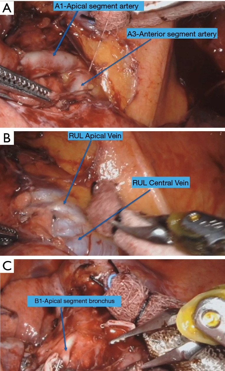
Right upper lobe apical segmentectomy (S1). (A) View of the first branch of right pulmonary artery dividing into anterior (A3) and apical branches (A1) with lung retracted caudally. (B) Right upper lobe vein with lung retracted posteriorly. (C) Right upper lobe apical segment bronchus (B1) after the transection of A1.
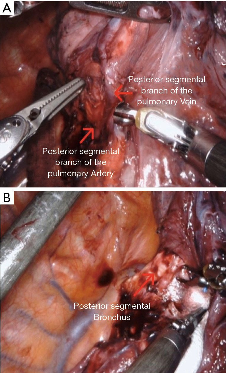
Right upper lobe posterior segmentectomy (S2). (A) Right upper lobe posterior segmental artery (A2) and Vein (V2). (B) Right upper lobe posterior segment bronchus (B2) after the transection of artery and vein.
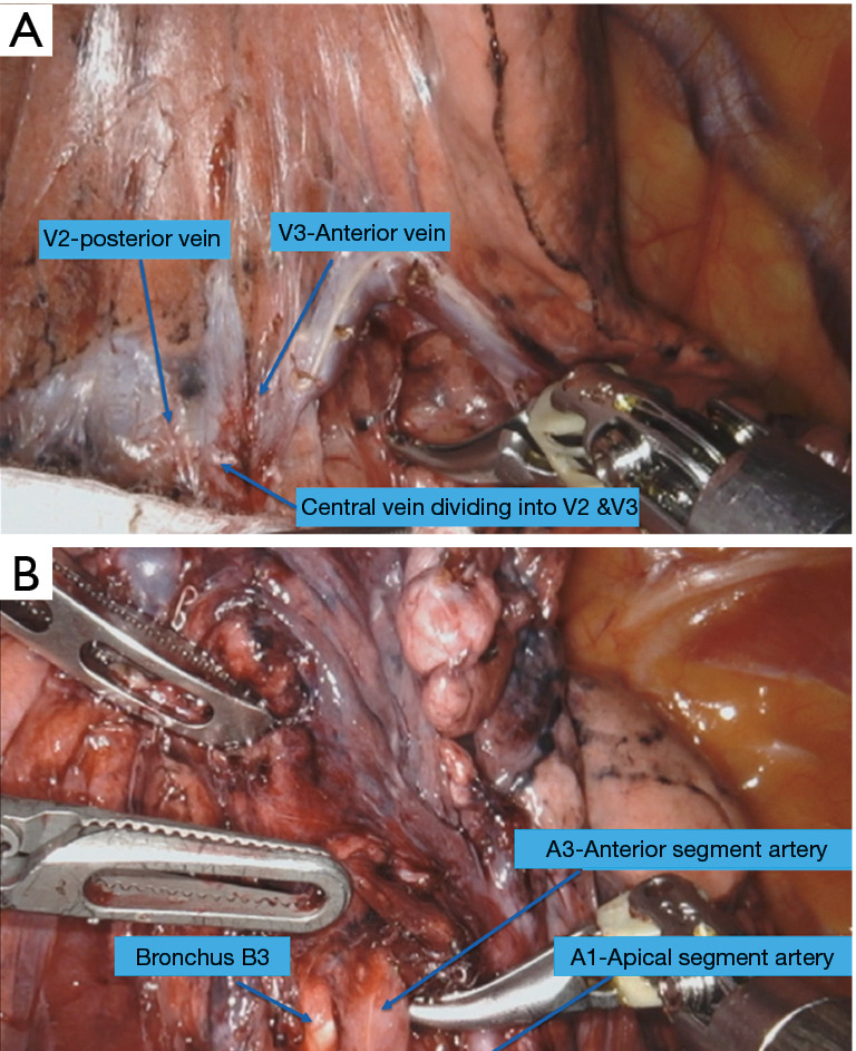
Right upper lobe anterior segmentectomy (S3). (A) Right upper lobe central vein division into posterior segmental (V2) and anterior segmental vein (V3). (B) Exposure of anterior segmental artery (A3) and bronchus (B3) after the division of the vein (V3).

Left upper lobe upper division segmentectomy (S1+S2+S3). (A) Left upper lobe apical and posterior segmental arteries. (B) Left upper lobe anterior segmental artery (A3) after the division of upper lobe upper division vein.
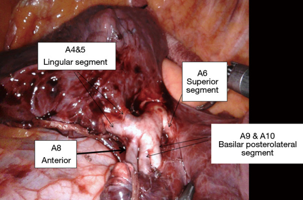
Left pulmonary artery in the fissure with exposure of lingular and lower lobe branches.
References
-
- National Cancer Institute: Surveillance, Epidemiology, and End Results Program. Reports on Cancer. [Internet]. April 2018. Available online: https://seer.cancer.gov/statfacts/html//common.html.
-
- De Koning H, Van Der Aalst C, Ten Haaf K, et al. Effects of volume CT lung cancer screening: mortality results of the Nelson randomised-controlled population based trial. Sept 2019. Presented at IASLC: 20th World Conference on Lung Cancer. Barcelona, Spain.
LinkOut - more resources
Full Text Sources