Review
. 2022 Jan;9(2):e2102678.
doi: 10.1002/advs.202102678. Epub 2021 Nov 19.

Mesoporous Bioactive Glasses in Cancer Diagnosis and Therapy: Stimuli-Responsive, Toxicity, Immunogenicity, and Clinical Translation

Affiliations
Review

Mesoporous Bioactive Glasses in Cancer Diagnosis and Therapy: Stimuli-Responsive, Toxicity, Immunogenicity, and Clinical Translation

Esmaeel Sharifi et al. Adv Sci (Weinh). 2022 Jan.

Abstract

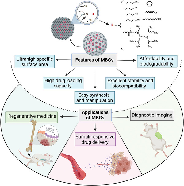
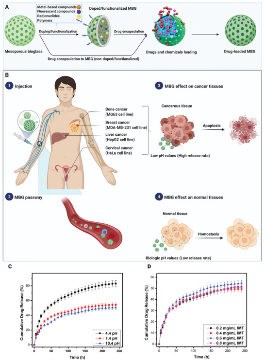
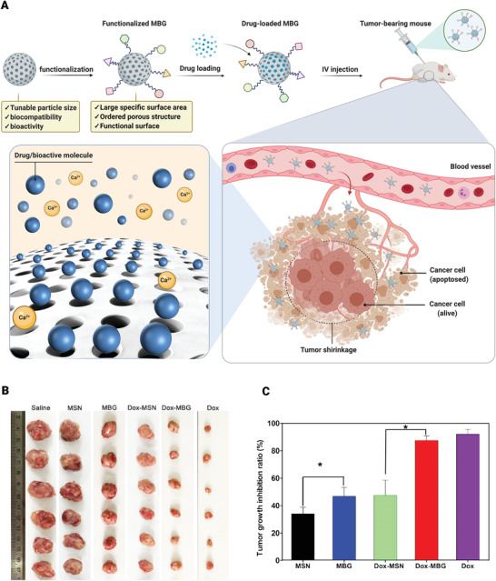
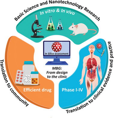
Cancer is one of the top life-threatening dangers to the human survival, accounting for over 10 million deaths per year. Bioactive glasses have developed dramatically since their discovery 50 years ago, with applications that include therapeutics as well as diagnostics. A new system within the bioactive glass family, mesoporous bioactive glasses (MBGs), has evolved into a multifunctional platform, thanks to MBGs easy-to-functionalize nature and tailorable textural properties-surface area, pore size, and pore volume. Although MBGs have yet to meet their potential in tumor treatment and imaging in practice, recently research has shed light on the distinguished MBGs capabilities as promising theranostic systems for cancer imaging and therapy. This review presents research progress in the field of MBG applications in cancer diagnosis and therapy, including synthesis of MBGs, mechanistic overview of MBGs application in tumor diagnosis and drug monitoring, applications of MBGs in cancer therapy ( particularly, targeted delivery and stimuli-responsive nanoplatforms), and immunological profile of MBG-based nanodevices in reference to the development of novel cancer therapeutics.

Keywords: cancer therapy; diagnosis; gene and drug delivery; immunotherapy; mesoporous bioactive glasses; toxicological profile.


Conflict of interest statement

The authors declare no conflict of interest.

Figures

Figure 1
Figure 1
An overview of mesoporous bioactive glass (MBG) properties and their potential biomedical applications.
Figure 2
Figure 2
Osteoblast and osteoclast cells' activity in healthy and cancerous bone plus bone cancer treatment through magnetic and light‐responsive materials. A) The Schematic indicates the normal and abnormal functions of osteoblast and osteoclast cells in cancerous bone cells.B) The schematic illustrates simultaneous bone regeneration and cancer therapy of stimuli‐responsive MBGs.
Figure 3
Figure 3
Schematic illustration of the MBGs synthesis by sol–gel process, EISA method, and bioinspired approach. CTAB: cetyltrimethylammonium bromide; HCl: hydrochloric acid; EISA: evaporation induced self‐assembly; Pluronic: an amphiphilic material based on poly(ethylene oxide)–poly(propylene oxide)–poly(ethylene oxide) (PEO–PPO–PEO) block copolymers.
Figure 4
Figure 4
Schematic illustration of the A) MBGs, B) drug‐loaded MBGs, and C) functionalized MBGs in diagnostic and drug delivery. CSCs: cancer stem cells.
Figure 5
Figure 5
Diagnosis application of bioactive glasses. A) Schematic illustration of the structure of UCNP@SiO2@MBG NPs. B) The luminescence in vivo imaging of athymic nude mice with intravenous injections of UCNPs@SiO2@MBG. Resolution/sensitivity of in vivo imaging enhanced by incorporating Ca (top row of in vivo imaging combined with Ca). All images were obtained under the same instrumental conditions (powder density ≈ 120 mW cm−2 on mice's surfaces). C) The loading capacity range of UCNPs@SiO2@MBG/ZnPc. D) MBG/UCNP nanocomposites' intensity versus release of ZnPc. E) Linear range of I/I o response for each release percentage. UCNPs: upconversion nanoparticles; ZnPc: zinc phthalocyanine. (B–E) Reproduced with permission.[ 93 ] Copyright 2016, Springer Nature.
Figure 6
Figure 6
Fabrication and applications of immunosensors based on bioactive glasses. A) Schematic of experimental pathways for preparation of electrochemical immunosensor and amperometric responses. B) TEM image of slit‐shaped PBG materials. C) Curve of amperometric responses and D) calibration curve of the prepared immunosensor for PCT detection with a wide linear range (500 fg mL−1 to 50 ng mL−1, Error bars or RSD for five measurements were calculated). MGCE: magnetic glass carbon electrode; PCT: procalcitonin; GA; glutaraldehyde; Ab1: the primary antibodies; PBG‐Ab2; the secondary antibodies‐pineal mesoporous bioactive glass. Reproduced with permission.[ 96 ] Copyright 2020, Elsevier.
Figure 7
Figure 7
A prospective overview of MBGs application as drug delivery platform. A) Preparation of drug‐loaded MBG. B) Drug‐loaded MBGs affect normal cells and various cancer cell lines. C) Drug release properties against different pH values. D) Drug release properties against different drug loading concentrations (IMT: Imatinib). (C,D) Reproduced with permission.[ 115 ] Copyright 2017, Elsevier.
Figure 8
Figure 8
MBGs application for anticancer drug delivery in vivo. A) Schematic illustration of MBG nanosphere functionalization mechanism and drug loading and drug‐loaded MBG effect on tumor size. B) In vivo antitumor efficacy of dendritic MBG nanospheres in a mouse tumor xenograft model. Mice were injected with saline, mesoporous silica nanoparticles (MSN), dendritic MBG, DOX–MSN, DOX–MBG, and free DOX. Image shows the solid tumors removed at the end of the study. C) Tumor growth inhibition ratio. (IV: Intravenous; *: p < 0.05). Reproduced with permission.[ 122 ] Copyright 2018, American Chemical Society.
Figure 9
Figure 9
Schematic of selective internal radiotherapy (SIRT) via 90Y‐MBG for hepatocellular carcinoma. A) Synthesis of yttrium‐loaded mesoporous bioactive glasses (90Y‐MBG). B) Application of 90Y‐MBG for radiotherapy. A catheter is passed through the femoral artery and guided to arteries supplying the liver. Then, 90Y‐MBG is infused through the catheter into the arteries. When they land in the tumor, radiotherapy is performed, and the emitted radiation kills the cancer cells.
Figure 10
Figure 10
The simultaneous suppression effect of Ca ions and DOX molecules on the tumor growth (pH = 6.5–6.8). Schematic illustration of the development and tumor suppression of the pH‐sensitive DOX‐loaded dendritic MBG step by step.
Figure 11
Figure 11
Multifunctional pH‐sensitive MBG for skin cancer therapy and regeneration. A) Schematic illustration of synthesis, decoration with folate‐alendronate (FAAL), and DOX loading of the mesoporous branched Eu‐Gd bioactive glass nanoparticles (EGBBGNs). The multifunctionality of EGBBGNs including imaging, melanoma therapy, and tissue regeneration. The interactions between the nanoparticles, surface modifiers, and DOX. B) Schematic on the inhibiting tumor recurrence. C) Photographs related to the wounds treated with different samples as follows: EGBBGNs‐FAAL (EG@F), EGBBGNs‐FAAL‐DOX (EG@F‐D), and F127 up to 14 days. D) The images related to the removed tumors of various samples. E) The release profiles of different samples at physiological and acidic media. Reproduced with permission.[ 144 ] Copyright 2021, Elsevier.
Figure 12
Figure 12
Eradication of cancerous tissues by different stimuli‐responsive MBGs. Schematic representation of magnetic‐mediated hyperthermia and photothermal and photodynamic therapy.
Figure 13
Figure 13
Treatment of a bone tumor through adopting magnetic‐responsive MBGs. The schematic shows how a magnetic‐responsive agent deals with remaining bone cancerous tissue after surgery.
Figure 14
Figure 14
The combination of photothermal and photodynamic therapies for bone cancer therapy and regeneration. A) A schematic showing how Mn‐doped MBG/C6 works for bone cancer therapy and regeneration. B) The heating curve of 5Mn‐doped MBG/C6 after being implanted into critical‐sized femoral bone defects of rats. C) The defects photographs of control (CTR) and 5Mn‐doped MBG/C6 groups after 8 weeks. D) The thermal images after applying 808 nm laser irradiation up to 20 min to the 5Mn‐doped MBG plus Van Gieson staining images of (a,b) CTR and (c,d) 5Mn‐doped MBG/C6 at 8 weeks; the short‐term photothermal therapy had no significant negative effect on the bone regeneration. M represents the Mn‐doped MBG‐Ce6 particles. (B–D) Reproduced with permission.[ 54 ] Copyright 2020, Elsevier.
Figure 15
Figure 15
Light‐responsive SrFe12O19/MBG/chitosan composite scaffold for simultaneous bone cancer therapy and regeneration in vivo. A) Micro‐CT images of defects after being implanted with different samples; blank control, mesoporous BG/chitosan (BCS), SrFe12O19–BG–chitosan (MBCS) at two different ratios of 1:3 (MBCS1:3) and 1:7 (MBCS1:7). B) Light images relating to the Van Giesons picrofuchsin‐stained sections of defects filled with the scaffolds up to 12 weeks. The new bone tissue and scaffolds can be seen in red and black, respectively. C) IR thermal images and D) temperature (°C) versus time (min) curves of the implanted MBCS1:3 scaffold into tumor‐bearing mice with and without laser irradiation. E) Fluorescence images of implanted MBCS1:3 scaffolds into tumor‐bearing mice with and without laser irradiation up to 12 days. F) The change in tumor size over time after being treated with the MBCS1:3 scaffolds, n = 5. Reproduced with permission.[ 172 ] Copyright 2018, Elsevier.
Figure 16
Figure 16
Protein corona formation and its determinant factors. A) Schematic illustration with a futuristic vision of the protein corona formation on MBG upon coming in contact with blood plasma. Reproduced with permission.[ 208 ] Copyright 2014, American Chemical Society. B) Nanoparticle and protein corona formation (adsorbed blue, green, and cyan globules) upon nanoparticles contact with a biological fluid. Reproduced with permission.[ 209 ] Copyright 2017, American Chemical Society. C) Diagram including major factors affecting protein corona formation; divided into three main categories. IV: Intravenous.
Figure 17
Figure 17
Extracellular and intracellular reactions due to structural changes of the protein corona. BG–protein interaction causes various signal modulations and toxic effects in biofluids and cells. Reversible and irreversible orientation and conformational changes of protein structure after adsorption can perturb downstream signaling that may be harmful to the host. Protein corona formation has different extracellular and intracellular effects. B) The pros and cons of protein corona formation. ER, endoplasmic reticulum; ROS, reactive oxygen species.[ 212 ]
Figure 18
Figure 18
Uptake of MBG nanoparticles by bone marrow‐derived dendritic cells (BMDCs). A) Flow cytometry analysis of unstimulated (green, US), or LPS (red) or Poly I:C (PI:C, blue) stimulated BMDCs treated or untreated (gray) with fluorescein isothiocyanate (FITC)‐labeled‐MBG nanoparticles during 24 h. Graphs show the percentage of FITC+ cells (middle panel) or the median of fluorescence intensity (MFI, right panel). Immature as well as mature BMDCs take up the nanosphere. B) Confocal microscopy analysis of BMDCs after incubation of 2 h with FITC‐MBG nanoparticles shows the cytosolic distribution of nanospheres. FITC‐MBG nanoparticles are shown in green, in blues is shown cell nucleus stained with Hoescht dye. C) Unstimulated, or LPS‐ or Poly I:C‐stimulated BMDCs were incubated (gray bars) or not (light blue bars) with nanospheres during 24 h and evaluated for the expression of CD11c, CD40, CD86, MHC II, CD80 surface markers. Graphs show the MFI. MBG nanoparticles do not alter the maturation status of BMDCs, except the expression of CD86. D) ELISA assay for the detection of IL‐6 in 24 h culture supernatants of BMDCs incubated (gray bars) or not (light blue bars) with nanospheres, in the presence or without stimuli. MBG nanoparticles do not induce the proinflammatory IL‐6 cytokine in immature BMDCs (US), indicating that these nanomaterials do not induce DC maturation by themselves. Reproduced under the terms of CC‐BY license open access.[ 229 ] Copyright 2020, MDPI.
Figure 19
Figure 19
A simplified view of activation and functional polarization of macrophages in response to MBG nanoparticles. A) Figure summarizes selected features of macrophages polarized toward the proinflammatory M1 or anti‐inflammatory/prohealing M2 phenotype. Depending on different microenvironmental cues, uncommitted (M0) macrophages undergo either classical (e.g., IFN‐γ + LPS or TNF) or alternative (e.g., IL‐4/IL‐13) activation, acquiring distinct phenotypic and functional properties. Cytokine release profile of M1‐polarized cells includes IL‐6, IL‐12, IL‐23, TNF‐α, IL‐1β, as well as NO and ROI. Cytokine release profile of M2‐polarized cells includes IL‐4, IL‐10, IL‐13, IL‐1ra. Induction and activation of M1 cells lead to inflammation and tissue damage, while M2‐polarized cells promote tissue repair and regeneration. B) Phagocytosis of MBG‐75S nanomaterials does not induce macrophage polarization toward the proinflammatory M1 phenotype. C) In vivo phagocytosis of Cu‐MBG nanoparticles by monocyte/macrophages leads to their activation and polarization into the anti‐inflammatory CD163+ M2 phenotype. IFN‐γ: interferon‐gamma; LPS: lipopolysaccharide; TNF‐α: tumor necrosis factor‐alpha; TLR: toll‐like receptor; TGF‐β: transforming growth factor‐beta; NO: nitric oxide; ROI: reactive oxygen intermediate.
Figure 20
Figure 20
Schematic illustration of some possible effects of ion‐incorporated MBGs for cancer therapy. Apoptosis can be induced by triggering mitochondrial damage and upregulating proapoptotic factors such as caspases. Furthermore, DNA damage and molecular pathways involved in cancer proliferation such as Akt, MAPK, and STAT3 can be modulated. As a major mechanism involved in cancer metastasis, angiogenesis of cancer cells can be regulated by MBGs by affecting HIF signaling pathway. Abbreviations: MBG, mesoporous bioactive glass; HIF, hypoxia‐inducible factor; TRP, transient receptor potential; CaSR, calcium‐sensing receptor; Cyt C, cytochrome C; STAT3, signal transducer and activator of transcription 3; Akt, protein kinase B; MAPK, mitogen‐activated protein kinase; DOX, doxorubicin.
Figure 21
Figure 21
The convergent pathway toward clinical translation of MBGs requires a necessary balance between in vitro, in vivo, and in silico studies.

Similar articles

Cited by

References

    1. Ashrafizadeh M., Najafi M., Makvandi P., Zarrabi A., Farkhondeh T., Samarghandian S., J. Cell. Physiol. 2020, 235, 9241. - PubMed
    1. Mohammadinejad R., Dehshahri A., Sagar Madamsetty V., Zahmatkeshan M., Tavakol S., Makvandi P., Khorsandi D., Pardakhty A., Ashrafizadeh M., Ghasemipour Afshar E., Zarrabi A., J. Controlled Release 2020, 325, 249. - PMC - PubMed
    1. Dabbagh Moghaddam F., Akbarzadeh I., Marzbankia E., Farid M., khaledi L., Reihani A. H., Javidfar M., Mortazavi P., Cancer Nanotechnol. 2021, 12, 14.
    1. Moghaddam F. D., Mortazavi P., Hamedi S., Nabiuni M., Roodbari N. H., Anticancer. Agents Med. Chem. 2020, 20, 790. - PubMed
    1. Makvandi P., Josic U., Delfi M., Pinelli F., Jahed V., Kaya E., Ashrafizadeh M., Zarepour A., Rossi F., Zarrabi A., Agarwal T., Zare E. N., Ghomi M., Kumar Maiti T., Breschi L., Tay F. R., Adv. Sci. 2021, 8, 2004014. - PMC - PubMed

Publication types

LinkOut - more resources