. 2022 Feb;33(2):326-341.
doi: 10.1681/ASN.2021050616. Epub 2021 Nov 19.

Xanthine Oxidoreductase Inhibitors Suppress the Onset of Exercise-Induced AKI in High HPRT Activity Urat1- Uox Double Knockout Mice

Affiliations

Xanthine Oxidoreductase Inhibitors Suppress the Onset of Exercise-Induced AKI in High HPRT Activity Urat1- Uox Double Knockout Mice

Takuji Hosoya et al. J Am Soc Nephrol. 2022 Feb.

Abstract

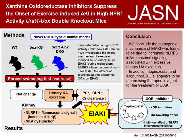
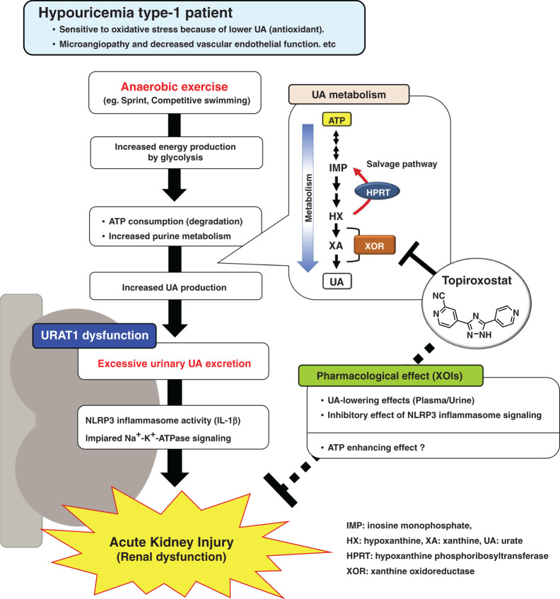
Background: Hereditary renal hypouricemia type 1 (RHUC1) is caused by URAT1/SLC22A12 dysfunction, resulting in urolithiasis and exercise-induced AKI (EIAKI). However, because there is no useful experimental RHUC1 animal model, the precise pathophysiologic mechanisms underlying EIAKI have yet to be elucidated. We established a high HPRT activity Urat1-Uox double knockout (DKO) mouse as a novel RHUC1 animal model for investigating the cause of EIAKI and the potential therapeutic effect of xanthine oxidoreductase inhibitors (XOIs).

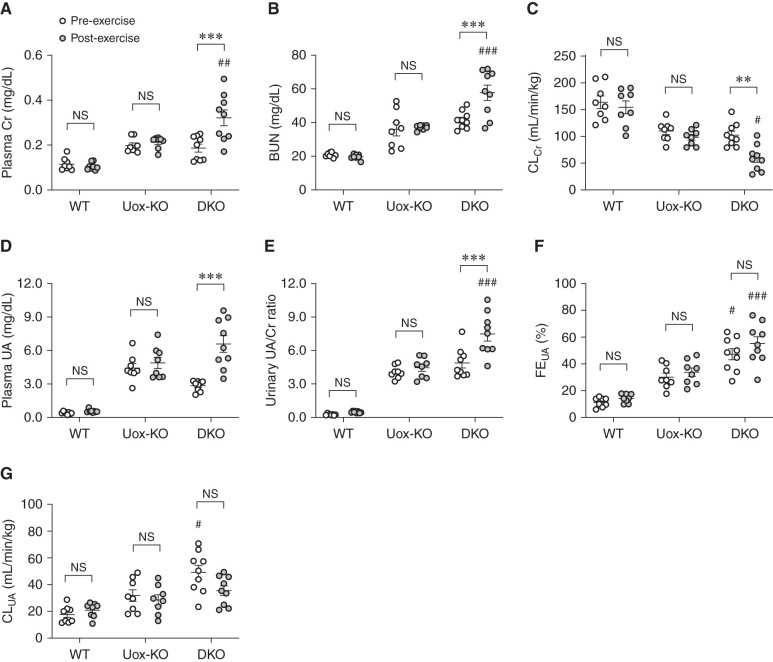
Methods: The novel Urat1-Uox DKO mice were used in a forced swimming test as loading exercise to explore the onset mechanism of EIAKI and evaluate related purine metabolism and renal injury parameters.

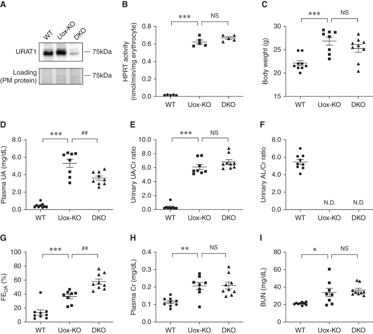
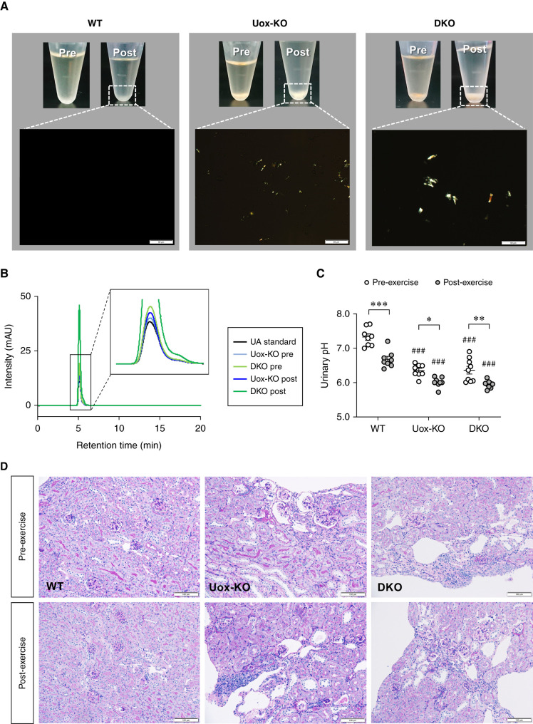
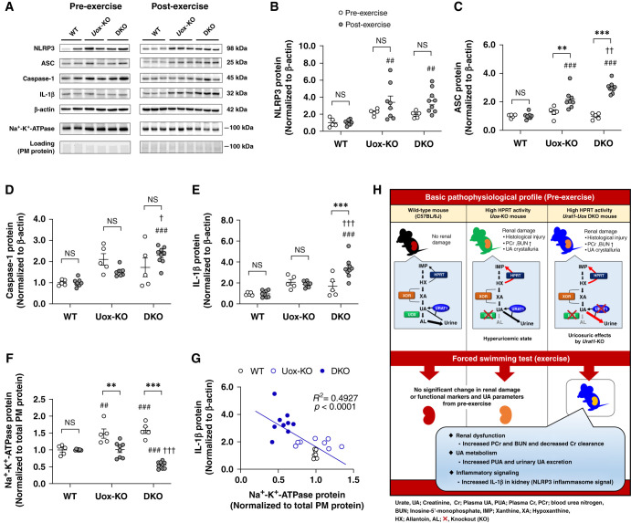
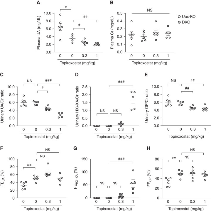
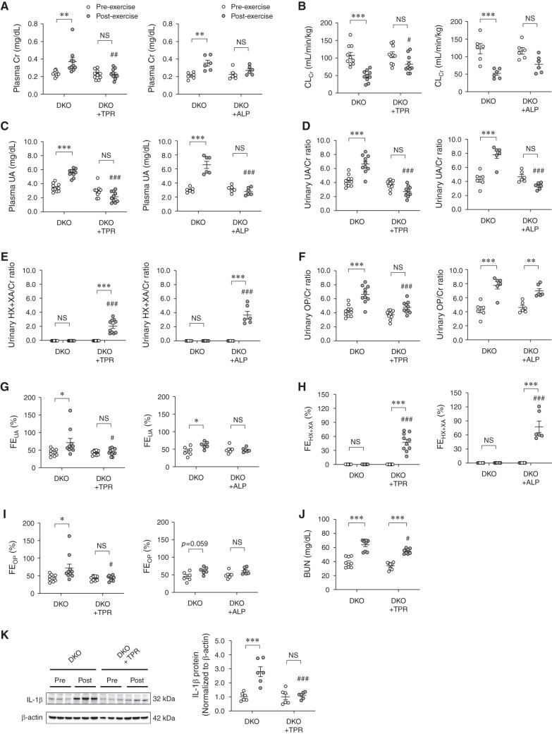
Results: Urat1-Uox DKO mice had uricosuric effects and elevated levels of plasma creatinine and BUN as renal injury markers, and decreased creatinine clearance observed in a forced swimming test. In addition, Urat1-Uox DKO mice had increased NLRP3 inflammasome activity and downregulated levels of Na+-K+-ATPase protein in the kidney, as Western blot analysis showed. Finally, we demonstrated that topiroxostat and allopurinol, XOIs, improved renal injury and functional parameters of EIAKI.

Conclusions: Urat1-Uox DKO mice are a useful experimental animal model for human RHUC1. The pathogenic mechanism of EIAKI was found to be due to increased levels of IL-1β via NLRP3 inflammasome signaling and Na+-K+-ATPase dysfunction associated with excessive urinary urate excretion. In addition, XOIs appear to be a promising therapeutic agent for the treatment of EIAKI.

Keywords: exercise-induced acute kidney injury (EIAKI); hypoxanthine phosphoribosyltransferase (HPRT); inborn errors; knockout; mice; renal hypouricemia (RHUC); renal tubular transport; urate transporter 1 (URAT1); urolithiasis; xanthine oxidoreductase inhibitor (XOI).


Figures

None
Graphical abstract
Figure 1.
Figure 1.
Establishment of male high HPRT activity Urat1-Uox DKO mice. (A) Expression of URAT1 protein in the kidney plasma membrane of male WT (C57BL/6J), high HPRT activity Uox-KO, and Urat1-Uox DKO mice (n=3 per group). (B) HPRT activity of mouse erythrocytes in male WT, high HPRT activity Uox-KO, and Urat1-Uox DKO mice (n=5 per group). (C) Body weight, (D) plasma UA, (E) urinary UA/Cr ratio, (F) urinary AL/Cr ratio, (G) FEUA, (H) plasma Cr, and (I) BUN in male WT (B6, n=9), high HPRT activity Uox-KO (n=8), and Urat1-Uox DKO mice (n=9). Data represent means±SEM; data were analyzed using 1-way ANOVA with Tukey’s post hoc test in (B–I). Significantly different from WT (*P<0.05, **P<0.01, ***P<0.001) and Uox-KO (##P<0.01). N.D., not detected; NS, not significant (P>0.05).
Figure 2.
Figure 2.
EIAKI of high HPRT activity Urat1-Uox DKO mice. (A) Plasma Cr, (B) BUN, (C) CLCr, (D) plasma UA, (E) urinary UA/Cr ratio, (F) FEUA, and (G) CLUA in WT (B6, n=8), high HPRT activity Uox-KO (n=8), and Urat1-Uox DKO mice (n=9) pre- and post-exercise. Data represent means±SEM; data were analyzed using 2-way ANOVA with Tukey’s post hoc test in (A–G). Significantly different from pre-exercise group (**P<0.01, ***P<0.001) and change in Uox-KO group (#P<0.05, ##P<0.01, ###P<0.001). NS, not significant (P>0.05).
Figure 3.
Figure 3.
Renal histology and UA crystalluria of high HPRT activity Urat1-Uox DKO mice. (A) Urine was collected from WT, Uox-KO, and Urat1-Uox DKO mice pre- and post-exercise. Bright signals in the lower images are urinary deposits. Urinary crystals were observed under polarized light (scale bar=50 μm). (B) A peak corresponding to UA was detected in solution after redissolving urinary crystals in HPLC buffer (refer to Supplemental Methods for details). (C) Urinary pH in WT (B6, n=8), Uox-KO (n=8), and Urat1-Uox DKO mice (n=9) pre- and post-exercise. (D) Periodic acid-Schiff staining of the kidney of WT, Uox-KO, and Urat1-Uox DKO mice pre- and post-exercise (scale bar =100 μm). Data represent means±SEM; data were analyzed using 2-way ANOVA with Tukey’s post hoc test in (C). Significantly different from pre-exercise group (*P<0.05, **P<0.01, ***P<0.001) and WT mice group (###P<0.001).
Figure 4.
Figure 4.
Expression of NLRP3 inflammasome related proteins and NKA alternated in kidney of high HPRT activity Urat1-Uox DKO mice pre- and post-exercise. (A) Representative Western blots of NLRP3, ASC, Caspase-1, IL-1β, β-actin, and NKA α1 in pre- and post-exercise. Protein expression of (B) NLRP3, (C) ASC, (D) Caspase-1, and (E) IL-1β in kidney of WT (pre, n=5; post, n=8), Uox-KO (pre, n=5; post, n=8), and Urat1-Uox DKO mice (pre, n=5; post, n=9) pre- and post-exercise as determined by Western blot analysis. Protein expression of (F) NKA α1 in the kidney plasma membrane-enriched (PM) protein of WT (pre, n=5; post, n=8), Uox-KO (pre, n=5; post, n=8), and Urat1-Uox DKO mice (pre, n=5; post, n=9) pre- and post-exercise as determined by Western blot analysis. (G) The correlation of IL-1β with NKA PM protein in kidney of WT, Uox-KO, and Urat1-Uox DKO mice after exercise. Dot plot graphs show the results of densitometric quantitation. Data represent means±SEM; data were analyzed using 2-way ANOVA with Tukey’s post hoc test in (B–F). Significantly different from pre-exercise group (**P<0.01, ***P<0.001), WT mice group (##P<0.01, ###P<0.001), and Uox-KO mice group (P<0.05, ††P<0.01, †††P<0.001). NS, not significant (P>0.05). (H) Schematic representation of the pathophysiologic features of model mice.
Figure 5.
Figure 5.
Effects of topiroxostat, a nonpurine-type XOI, on purine metabolism of high HPRT activity Urat1-Uox DKO mice. (A) Plasma UA, (B) plasma Cr, (C) urinary UA/Cr ratio, (D) urinary HX+XA/Cr ratio, (E) urinary OP/Cr ratio, (F) FEUA, (G) FEHX+XA, and (H) FEOP in high HPRT activity Uox-KO (n=6) and Urat1-Uox DKO mice (DKO control, n=5; topiroxostat 0.3 mg/kg, n=6; topiroxostat 1 mg/kg, n=5). Data represent means±SEM; data were analyzed using unpaired t test (Uox-KO versus DKO control) and 1-way ANOVA with Dunnett’s post hoc test (DKO control versus topiroxostat-treated groups) in (A–H). Significantly different from Uox-KO group (*P<0.05, **P<0.01) and Urat1-Uox DKO group (#P<0.05, ##P<0.01, ###P<0.001). NS, not significant (P>0.05).
Figure 6.
Figure 6.
Effects of topiroxostat and allopurinol, XOIs, on EIAKI of high HPRT activity Urat1-Uox DKO mice. (A) Plasma Cr, (B) CLCr, (C) plasma UA, (D) urinary UA/Cr ratio, (E) urinary HX+XA/Cr ratio, (F) urinary OP/Cr ratio, (G) FEUA, (H) FEHX+XA, and (I) FEOP in Urat1-Uox DKO control group (DKO, n=10) and topiroxostat-treated Urat1-Uox DKO group (DKO+TPR, n=10) or Urat1-Uox DKO control group (DKO, n=6) and allopurinol-treated Urat1-Uox DKO group (DKO+ALP, n=6) pre- and post-exercise. (J) BUN in Urat1-Uox DKO control group (DKO, n=10) and topiroxostat-treated Urat1-Uox DKO group (DKO+TPR, n=10) pre- and post-exercise. (K) IL-1β in the kidney of the Urat1-Uox DKO control group (DKO, n=6) and topiroxostat-treated Urat1-Uox DKO group (DKO+TPR, n=6) pre- and post-exercise as determined by Western blot analysis. Data represent means±SEM; data were analyzed using 2-way ANOVA with Tukey’s post hoc test in (A–K). Significantly different from pre-exercise groups (*P<0.05, **P<0.01, ***P<0.001) and Urat1-Uox DKO control group (#P<0.05, ##P<0.01, ###P<0.001). NS, not significant (P>0.05). ALP, allopurinol; TPR, topiroxostat.
Figure 7.
Figure 7.
Schematic representation of the mechanism of EIAKI on RHUC1 together with the therapeutic action of XOIs.

References

    1. Nakayama A, Matsuo H, Ohtahara A, Ogino K, Hakoda M, Hamada T, et al. : Clinical practice guideline for renal hypouricemia (1st edition). Hum Cell 32: 83–87, 2019 - PMC - PubMed
    1. Enomoto A, Kimura H, Chairoungdua A, Shigeta Y, Jutabha P, Cha SH, et al. : Molecular identification of a renal urate anion exchanger that regulates blood urate levels. Nature 417: 447–452, 2002 - PubMed
    1. Ichida K, Hosoyamada M, Hisatome I, Enomoto A, Hikita M, Endou H, et al. : Clinical and molecular analysis of patients with renal hypouricemia in Japan-influence of URAT1 gene on urinary urate excretion. J Am Soc Nephrol 15: 164–173, 2004 - PubMed
    1. Ichida K, Hosoyamada M, Kamatani N, Kamitsuji S, Hisatome I, Shibasaki T, et al. : Age and origin of the G774A mutation in SLC22A12 causing renal hypouricemia in Japanese. Clin Genet 74: 243–251, 2008 - PubMed
    1. Vidanapathirana DM, Jayasena S, Jasinge E, Stiburkova B: A heterozygous variant in the SLC22A12 gene in a Sri Lanka family associated with mild renal hypouricemia. BMC Pediatr 18: 210, 2018 - PMC - PubMed

Publication types

MeSH terms

Supplementary concepts

LinkOut - more resources