. 2021 Nov 19;11(1):22594.
doi: 10.1038/s41598-021-01963-3.

Cuprizone feed formulation influences the extent of demyelinating disease pathology

Affiliations

Cuprizone feed formulation influences the extent of demyelinating disease pathology

Lillian M Toomey et al. Sci Rep. .

Abstract

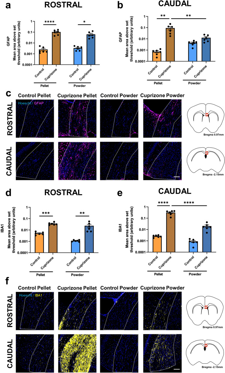
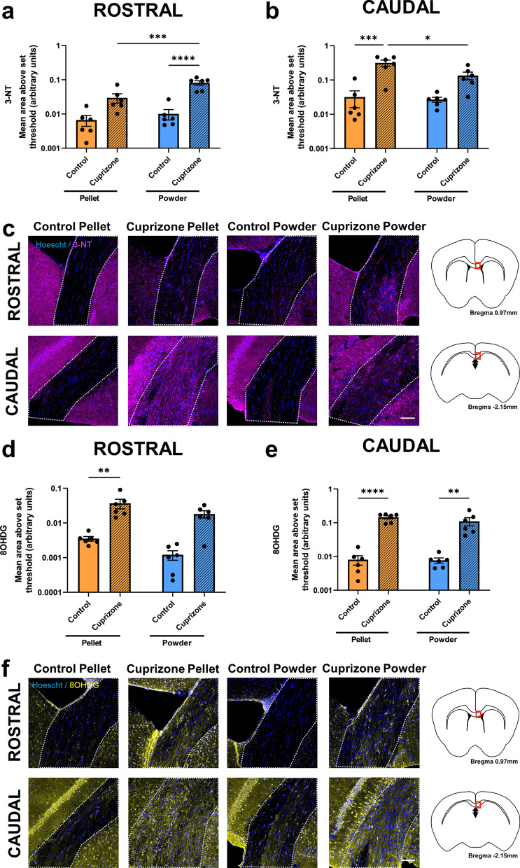
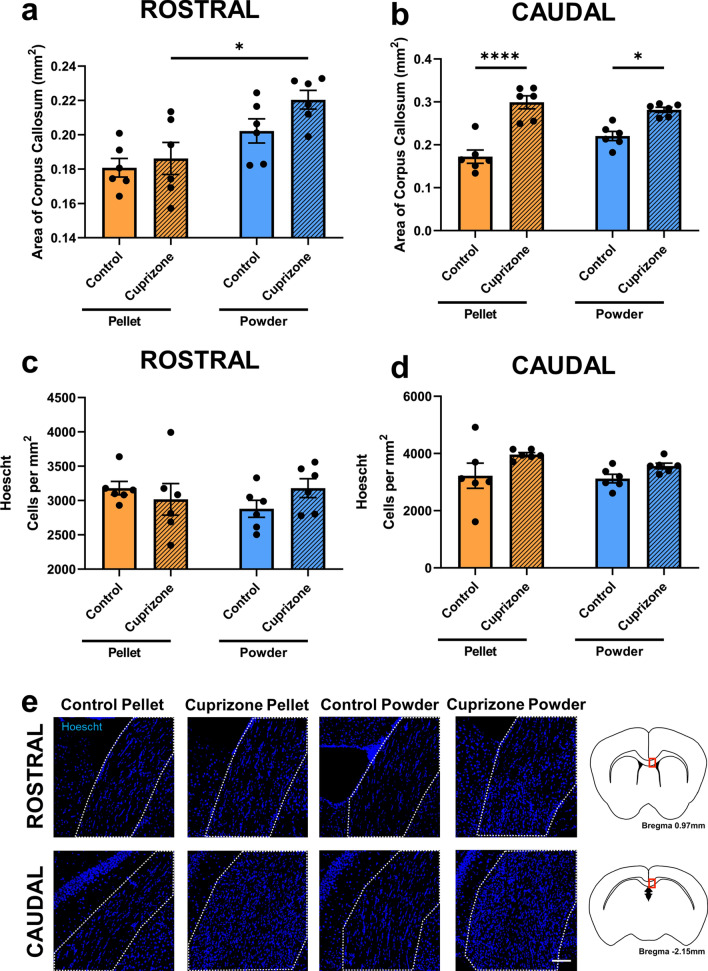
Cuprizone is a copper-chelating agent that induces pathology similar to that within some multiple sclerosis (MS) lesions. The reliability and reproducibility of cuprizone for inducing demyelinating disease pathology depends on the animals ingesting consistent doses of cuprizone. Cuprizone-containing pelleted feed is a convenient way of delivering cuprizone, but the efficacy of these pellets at inducing demyelination has been questioned. This study compared the degree of demyelinating disease pathology between mice fed cuprizone delivered in pellets to mice fed a powdered cuprizone formulation at an early 3 week demyelinating timepoint. Within rostral corpus callosum, cuprizone pellets were more effective than cuprizone powder at increasing astrogliosis, microglial activation, DNA damage, and decreasing the density of mature oligodendrocytes. However, cuprizone powder demonstrated greater protein nitration relative to controls. Furthermore, mice fed control powder had significantly fewer mature oligodendrocytes than those fed control pellets. In caudal corpus callosum, cuprizone pellets performed better than cuprizone powder relative to controls at increasing astrogliosis, microglial activation, protein nitration, DNA damage, tissue swelling, and reducing the density of mature oligodendrocytes. Importantly, only cuprizone pellets induced detectable demyelination compared to controls. The two feeds had similar effects on oligodendrocyte precursor cell (OPC) dynamics. Taken together, these data suggest that demyelinating disease pathology is modelled more effectively with cuprizone pellets than powder at 3 weeks. Combined with the added convenience, cuprizone pellets are a suitable choice for inducing early demyelinating disease pathology.


Conflict of interest statement

The authors declare no competing interests.

Figures

Figure 1
Figure 1
Effects of cuprizone feed formulation on animal weights. Animals were weighed daily to assess weight loss throughout cuprizone administration. Cuprizone was delivered in either pelleted or powdered form. N = 6 mice per group; graph displays mean weight ± SEM. Significant differences are indicated by *p ≤ 0.05, **p ≤ 0.01.
Figure 2
Figure 2
Effects of cuprizone feed formulation on glial reactivity. Area of immunointensity of GFAP in the rostral (a) and caudal (b) corpus callosum was assessed to determine the level of astrocyte reactivity. Area of immunointensity of IBA1 in the rostral (d) and caudal (e) corpus callosum was assessed to determine the level of microglial activation. N = 6 mice per group, graphs display individual data points overlayed on a bar displaying the mean ± SEM. Significant differences are indicated by *p ≤ 0.05, **p ≤ 0.01, ***p ≤ 0.001, ****p ≤ 0.0001. Representative images of the area of GFAP (c) and IBA1 (f) immunoreactivity is shown, scale bars = 100 µm. Area of the corpus callosum analysed is denoted by dotted lines. This area is indicated by the red box in the coronal overviews illustrating the rostral and caudal regions of the corpus callosum (c,f). The stereotaxic coordinates are indicated.
Figure 3
Figure 3
Effects of cuprizone feed formulation on oxidative stress. Area of immunointensity of 3-NT in the rostral (a) and caudal (b) corpus callosum was assessed to determine the level of protein nitration. Area of immunointensity of 8OHDG in the rostral (d) and caudal (e) corpus callosum was assessed to determine the level of oxidative DNA damage. N = 6 mice per group, graphs display individual data points overlayed on a bar displaying the mean ± SEM. Significant differences are indicated by *p ≤ 0.05, **p ≤ 0.01, ***p ≤ 0.001, ****p ≤ 0.0001. Representative images of the area of 3-NT (c) and 8OHDG (f) immunoreactivity are shown, scale bars = 100 µm. Area of the corpus callosum analysed is denoted by dotted lines. This area is indicated by the red box in the coronal overviews illustrating the rostral and caudal regions of the corpus callosum (c,f). The stereotaxic coordinates are indicated.
Figure 4
Figure 4
Effects of cuprizone feed formulation on tissue swelling. The area of the corpus callosum in the rostral (a) and caudal (b) areas was assessed. The density of Hoechst+ cells was also quantified in the rostral (c) and caudal (d) corpus callosum. N = 6 mice per group, graphs display individual data points overlayed on a bar displaying the mean ± SEM. Significant differences are indicated by *p ≤ 0.05, **p ≤ 0.01, ***p ≤ 0.001, ****p ≤ 0.0001. Representative images Hoechst+ cells (e) is shown, scale bars = 100 µm. Area of the corpus callosum analysed is denoted by dotted lines. This area is indicated by the red box in the coronal overviews illustrating the rostral and caudal regions of the corpus callosum (e). The stereotaxic coordinates are indicated.
Figure 5
Figure 5
Effects of cuprizone feed formulation on densities of Olig2+, PDGFRα+ and Olig2+/PDGFRα+ cells. The area of the rostral and caudal corpus callosum analysed is denoted by the red box in the coronal illustrations of the brain (a,b). The density of Olig2+ cells (c,d), PDGFRα+ cells (e,f) and Olig2+/PDGFRα+ cells (g,h) were quantified in the rostral and caudal corpus callosum. N = 6 mice per group, graphs display individual data points overlayed on a bar displaying the mean ± SEM. Significant differences are indicated by *p ≤ 0.05, **p ≤ 0.01, ***p ≤ 0.001, ****p ≤ 0.0001. (i) Representative images of Olig2+, PDGFRα+ and Olig2+/PDGFRα+ cells, indicated with arrow heads; scale bar = 25 µm.
Figure 6
Figure 6
Effects of cuprizone feed formulation on densities of ASPA+ and CC1+ cells. The area of the rostral and caudal corpus callosum analysed is denoted by the red box in the coronal illustrations of the brain (a,b). The density of ASPA+ cells (c,d) and CC1+ cells (e,f) were quantified in the rostral and caudal corpus callosum. N = 6 mice per group, graphs display individual data points overlayed on a bar displaying the mean ± SEM. Significant differences are indicated by *p ≤ 0.05, **p ≤ 0.01, ***p ≤ 0.001, ****p ≤ 0.0001. (g) Representative images of ASPA+ and CC1+ cells, indicated with white arrow heads; example ASPA-/CC1+ cell indicated with yellow arrow head; scale bar = 25 µm.
Figure 7
Figure 7
Effects of cuprizone feed formulation on demyelination. The area of the rostral and caudal corpus callosum analysed is denoted by the red box in the coronal illustrations of the brain (a,b). Percentage area of staining of Black-Gold II in the rostral (c) and caudal (d) corpus callosum was assessed to determine the level of myelination. N = 6 mice per group, graphs display individual data points overlayed on a bar displaying the mean ± SEM. Significant differences are indicated by *p ≤ 0.05, **p ≤ 0.01, ***p ≤ 0.001, ****p ≤ 0.0001. (e) Representative images of the area of Black-Gold II for each group is shown, 4 × images scale bar = 500 µm; 10 × images scale bar = 200 µm.

References

    1. Matsushima GK, Morell P. The neurotoxicant, cuprizone, as a model to study demyelination and remyelination in the central nervous system. Brain Pathol. 2001;11:107–116. - PMC - PubMed
    1. Kipp M, et al. The cuprizone animal model: New insights into an old story. Acta Neuropathol. 2009;118:723–736. - PubMed
    1. Torkildsen Ø, Brunborg LA, Myhr KM, Bø L. The cuprizone model for demyelination. Acta Neurol. Scand. 2008;117:72–76. - PubMed
    1. Praet J, Guglielmetti C, Berneman Z, Van der Linden A, Ponsaerts P. Cellular and molecular neuropathology of the cuprizone mouse model: Clinical relevance for multiple sclerosis. Neurosci. Biobehav. Rev. 2014;47:485–505. - PubMed
    1. Zhang Y, et al. Quetiapine alleviates the cuprizone-induced white matter pathology in the brain of C57BL/6 mouse. Schizophr. Res. 2008;106:182–191. - PubMed

Publication types