. 2021 Nov 19;4(1):1309.
doi: 10.1038/s42003-021-02787-7.

Allelic variants of full-length VAR2CSA, the placental malaria vaccine candidate, differ in antigenicity and receptor binding affinity

Affiliations

Allelic variants of full-length VAR2CSA, the placental malaria vaccine candidate, differ in antigenicity and receptor binding affinity

Jonathan P Renn et al. Commun Biol. .

Erratum in

Abstract

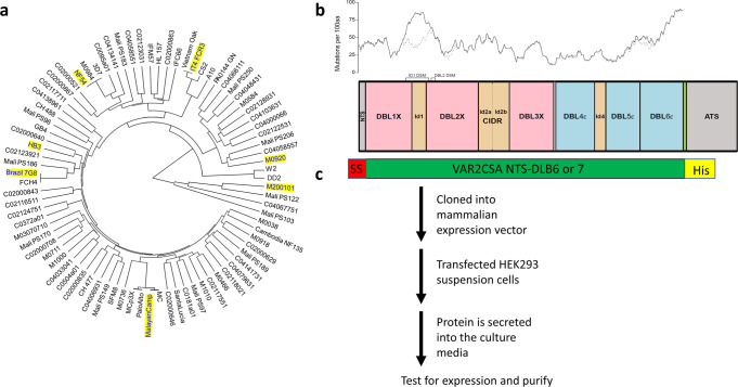
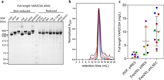
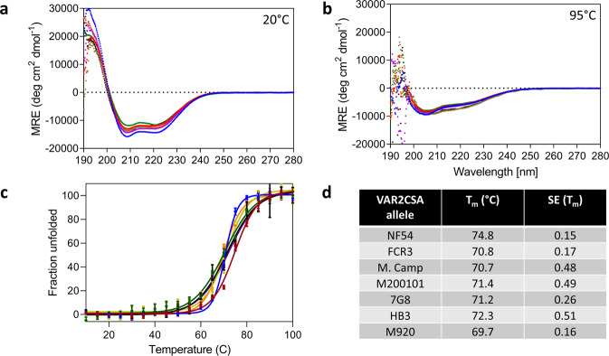
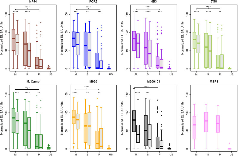
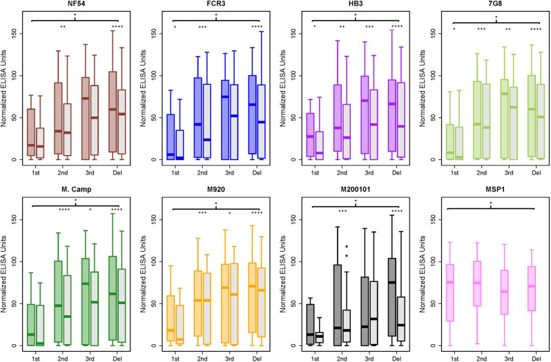
Plasmodium falciparum-infected erythrocytes (IE) sequester in the placenta via surface protein VAR2CSA, which binds chondroitin sulfate A (CSA) expressed on the syncytiotrophoblast surface, causing placental malaria (PM) and severe adverse outcomes in mothers and their offspring. VAR2CSA belongs to the PfEMP1 variant surface antigen family; PfEMP1 proteins mediate IE adhesion and facilitate parasite immunoevasion through antigenic variation. Here we produced deglycosylated (native-like) and glycosylated versions of seven recombinant full-length VAR2CSA ectodomains and compared them for antigenicity and adhesiveness. All VAR2CSA recombinants bound CSA with nanomolar affinity, and plasma from Malian pregnant women demonstrated antigen-specific reactivity that increased with gravidity and trimester. However, allelic and glycosylation variants differed in their affinity to CSA and their serum reactivities. Deglycosylated proteins (native-like) showed higher CSA affinity than glycosylated proteins for all variants except NF54. Further, the gravidity-related increase in serum VAR2CSA reactivity (correlates with acquisition of protective immunity) was absent with the deglycosylated form of atypical M200101 VAR2CSA with an extended C-terminal region. Our findings indicate significant inter-allelic differences in adhesion and seroreactivity that may contribute to the heterogeneity of clinical presentations, which could have implications for vaccine design.


Conflict of interest statement

The authors declare no competing interests as defined by Nature Research, or other interests that might be perceived to influence the results and/or discussion reported in this paper.

Figures

Fig. 1
Fig. 1. Phylogenetic analysis of VAR2CSA sequences from different isolates, frequency of amino acid mutations across VAR2CSA extracellular domains for the seven VAR2CSA alleles studied, and workflow of VAR2CSA expression.
a Phylogenetic tree showing full-length VAR2CSA amino acid sequences from lab strains and isolates from different geographic locations. The seven sequences selected for this study are highlighted in yellow. b Schematic organization of VAR2CSA protein for FCR3, drawn to scale, showing its N-terminal sequence, 6 cysteine-rich Duffy binding like (DBL) domains, inter-domain (ID) regions, transmembrane domain (TMD), and intracytoplasmic tail (ATS). Prevalence of sequence variability along the protein’s extent, using DBL1-6 from the seven full-length constructs (DBL7 from M20010 is not included in this analysis), is graphically displayed as a solid black line showing the number of amino acid mutations (not fully conserved in all seven constructs) per 100 consecutive amino acids, measured as a moving average inside a 100aa window stepping along the sequence. The locations of DSMs are highlighted. The dotted black line shows the impact of omitting one isolate M920 having the ID1DSM. c A schematic representation of the construct used for protein expression and the workflow for protein production. The extracellular domain of VAR2CSA (NTS-DBL6 or -DLB7, see Table 1 for exact boundaries) was cloned into a mammalian expression vector flanked by an N-terminal signal sequence (red) that directs the protein for export and a C-terminal his tag (yellow) for detection and purification.
Fig. 2
Fig. 2. Expression and purification of full-length VAR2CSA, including SDS-PAGE analysis and protein yields.
a VAR2CSA recombinants expressed in Expi293 cells were analyzed in SDS-PAGE gels. Ten µg of purified full-length VAR2CSA FCR3 (lane 1 and 9), NF54 (lane 2 and 10), 7G8 (lane 3 and 11), HB3 (lane 4 and 12) M. Camp (lane 5 and 13), M920 (lane 6 and 14), and M200101 (lane 7 and 15) were loaded on a 3–8% tris-acetate gel (with or without 100 mM DTT) and Coomassie-stained. Lane 8 is the molecular weight marker (kDa). The theoretical sizes of full-length VAR2CSA range from 308 kDa to 346 kDa. b Size exclusion analysis of all 7 full-length VAR2CSA proteins ran on a Superose 6 column. Each trace is in a different color (FCR3 = blue, NF54 = red, HB3 = purple, 7G8 = yellow, M. Camp = green, M920-orange, M200101 = black). All proteins appear predominantly as a single species that elute around 14 mL (void volume of the column is 8 mL). c Different HEK293 suspension cell lines were combined with different expression plasmids. Protein yields for each allele is represented in a different color dot. All the preps ranged from 0.5 to 2L of transfected cells. The average yield of 293F plus pIRES plasmid was 1.4 mg/L ± 0.9, the average yield of Expi293 plus pIRES was 6.6 mg/L ± 5.2, and the average yield of Expi293 plus pHLSEC plasmid was 9.3 mg/L ± 5.9. Error is the standard deviation based on 7, 6, and 11 preps respectively.
Fig. 3
Fig. 3. Biophysical characterization of full-length VAR2CSA using circular dichroism spectroscopy.
Far‐UV CD wavelength scan of 0.2 mg/mL full-length VAR2CSA (FCR3 = blue, NF54 = red, HB3 = purple, 7G8 = yellow, M. Camp = green, M920-orange, M200101 = black) at a 20 °C and b 95 °C, showing the loss of α-helical structure upon thermal denaturation. c Thermal melt of all the seven alleles monitoring the loss of CD signal at 200 nm and plotting the fraction unfolded vs. temperature. d The Tm values of all seven alleles obtained by the fit. NF54 is statistically different. Error is SEM based on three biological replicates.
Fig. 4
Fig. 4. Full-length VAR2CSA recombinants are recognized by naturally acquired antibodies in gravidity- and glycosylation-dependent manner.
ELISA plates were coated with full-length VAR2CSA 150 samples from malaria-exposed multigravid (MG), secundigravid (SG), and primigravid (PG) were diluted 1:1000 and measured for reactivity to all full-length VAR2CSA proteins. Box and Whisker representation of ELISA reactivity to VAR2CSA from 150 samples from malaria-exposed women stratified by gravidity (n = 50 multigravida, n = 50 secundigravida and n = 50 primigravida) and 50 US naive samples. The solid line represents the median of reactivity, error bars represent 1.5 times the interquartile range. For each allele (FCR3 = blue, NF54 = red, HB3 = purple, 7G8 = yellow, M. Camp = green, M920-orange, M200101 = black), the glycosylated protein shaded in the corresponding color and the deglycosylated is shaded in gray. The most highly significant difference between MG and PG is displayed with the bracket indicated by **** (p < 0.0001); significant differences between MG and PG were identical for both glycosylated and deglycosylated proteins (for a more detailed analysis between the groups see Supplementary Figs. S5 and S6). Statistically significant differences between the values of the glycosylated and deglycosylated are indicated with *p < 0.05, **p < 0.01, ***p < 0.001, and ****p < 0.0001. The p-values for both comparisons were generated by the unpaired Mann–Whitney test.
Fig. 5
Fig. 5. Recognition of full-length VAR2CSA recombinants by naturally acquired antibodies increases with trimester.
The same data presented in Fig. 4 were separated into different bins of trimester instead of gravidity. The trimester bins were defined as first (≤13 weeks gestation), second (14–26 weeks), third (≥27 weeks), and time of delivery. Box and Whisker representation of ELISA reactivity to VAR2CSA from 148 samples stratified by trimester (n = 11 first, n = 46 second, n = 20 third, n = 71 delivery) from malaria-exposed women and 50 US naive samples. The solid line represents the median of reactivity, error bars represent 1.5 times the interquartile range. For each allele (FCR3 = blue, NF54 = red, HB3 = purple, 7G8 = yellow, M. Camp=green, M920-orange, M200101 = black), the glycosylated protein shaded in the corresponding color and the deglycosylated is shaded in gray. The significant difference between the first trimester and delivery for the glycosylated protein is displayed with the bracket indicated by * (p < 0.05). After deglycosylation, significant differences (p < 0.05) between first trimester and delivery remained for each allele except for NF54 (for a more detailed analysis see Supplementary Figs. S7 and S8). Statistically significant differences between the values of the glycosylated and deglycosylated are indicated with *p < 0.05, **p<0.01, ***p < 0.001, and ****p < 0.0001. The p-values for both comparisons were generated by the unpaired Mann–Whitney test.

References

    1. Desai M, et al. Epidemiology and burden of malaria in pregnancy. Lancet Infect. Dis. 2007;7:93–104. doi: 10.1016/S1473-3099(07)70021-X. - DOI - PubMed
    1. McGready R, Nosten F, Barnes KI, Mokuolu O, White NJ. Why is WHO failing women with falciparum malaria in the first trimester of pregnancy? Lancet. 2020;395:779. doi: 10.1016/S0140-6736(20)30161-6. - DOI - PubMed
    1. Langhorne J, Ndungu FM, Sponaas AM, Marsh K. Immunity to malaria: more questions than answers. Nat. Immunol. 2008;9:725–732. doi: 10.1038/ni.f.205. - DOI - PubMed
    1. Marsh K, Otoo L, Hayes RJ, Carson DC, Greenwood BM. Antibodies to blood stage antigens of Plasmodium falciparum in rural Gambians and their relation to protection against infection. Trans. R. Soc. Trop. Med Hyg. 1989;83:293–303. doi: 10.1016/0035-9203(89)90478-1. - DOI - PubMed
    1. Brabin BJ. An analysis of malaria in pregnancy in Africa. Bull. World Health Organ. 1983;61:1005–1016. - PMC - PubMed

Publication types

Substances