. 2022 Apr;29(4):818-831.
doi: 10.1038/s41418-021-00897-7. Epub 2021 Nov 19.

The Smad4-MYO18A-PP1A complex regulates β-catenin phosphorylation and pemigatinib resistance by inhibiting PAK1 in cholangiocarcinoma

Affiliations

The Smad4-MYO18A-PP1A complex regulates β-catenin phosphorylation and pemigatinib resistance by inhibiting PAK1 in cholangiocarcinoma

Jialiang Liu et al. Cell Death Differ. 2022 Apr.

Abstract

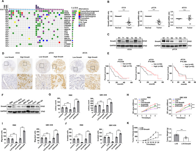
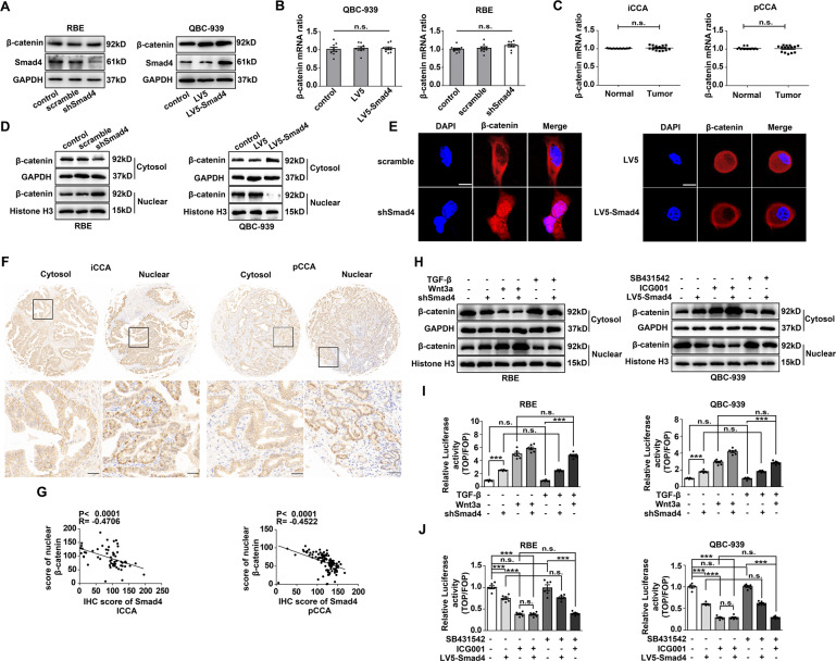
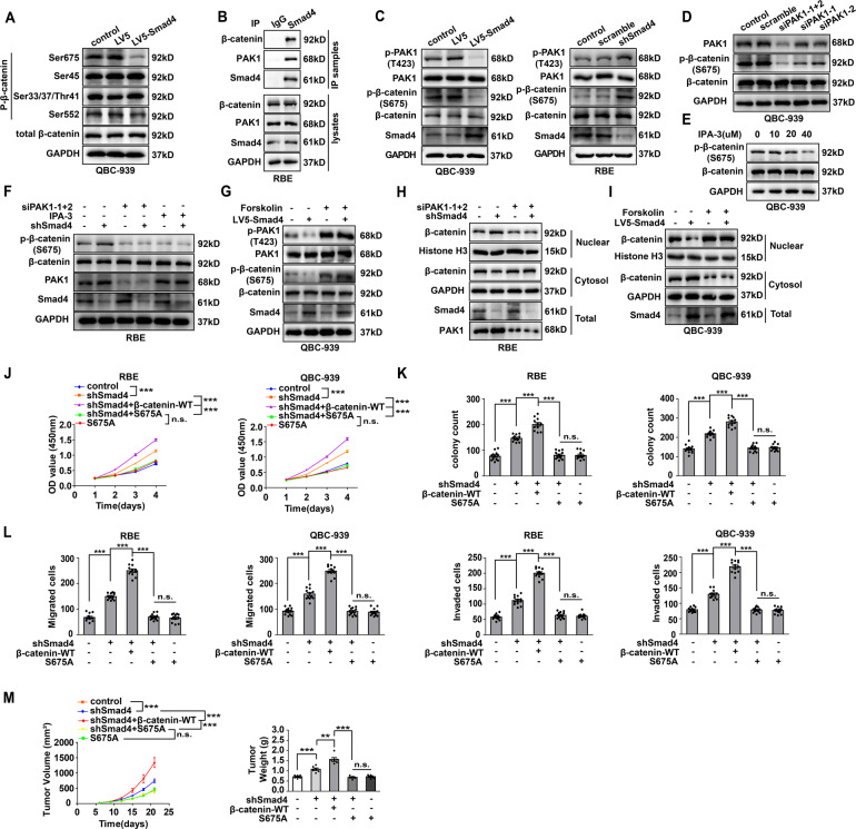
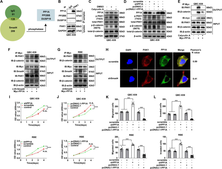
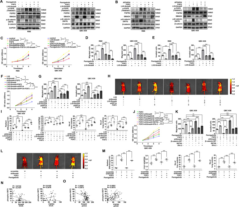
Cholangiocarcinoma (CCA), consisting of three subtypes-intrahepatic (iCCA), perihilar (pCCA), and distal (dCCA), is a highly aggressive cancer arising from the bile duct and has an extremely poor prognosis. Pemigatinib is the only FDA-approved targeted drug for CCA, and the CCA treatment options are substantially insufficient considering its poor prognosis and increasing morbidity. Here, we performed next-generation sequencing (NGS) of 15 pCCAs and 16 dCCAs and detected the expression of SMAD4, a frequently mutated gene, in 261 CCAs. By univariate and multivariate analyses, we identified Smad4 as a favorable prognostic biomarker in iCCA and pCCA. With in vitro and in vivo experiments, we demonstrated that Smad4 suppressed CCA proliferation, migration and invasion by inhibiting β-catenin-S675 phosphorylation and intranuclear translocation. We applied LC-MS/MS and multiple biochemical techniques and identified PP1A as the phosphatase in Smad4-mediated dephosphorylation of PAK1-T423, which is responsible for β-catenin-S675 phosphorylation. Moreover, we demonstrated that MYO18A is the PP1-interacting protein of PP1A for substrate recognition in CCA. MYO18A interacts with PP1A via its RVFFR motif and interacts with Smad4 via CC domain. Patients with coexpression of MYO18A and Smad4 have a more favorable prognosis than other patients. Smad4 enhances Pemigatinib efficiency, and Smad4 knockdown results in Pemigatinib resistance. In conclusion, coexpression of Smad4 and MYO18A is a favorable prognostic indicator for iCCA and pCCA. The Smad4-MYO18A-PP1A complex dephosphorylates PAK1-T423 and thus inhibits β-catenin-S675 phosphorylation and its intranuclear localization. Smad4 suppresses CCA proliferation, migration, invasion, and sensitivity to Pemigatinib by governing the phosphorylation and intracellular localization of β-catenin.


Conflict of interest statement

The authors declare no competing interests.

Figures

Fig. 1
Fig. 1. The genomic alteration, expression and function of Smad4 in CCA.
A Frequency of genomic alterations in 15 pCCAs and 16 dCCAs. B, C The expression of Smad4 was detected with qPCR in 15 pairs of CCAs and adjacent tissues (B), and with WB in three pairs of CCAs and adjacent tissues (C). D Smad4 expression was detected by IHC, and patients with CCA were divided into subsets with low and high Smad4 expression. Scale bar: 50 μm. E The correlation between Smad4 expression and overall survival in CCA was calculated with the log-rank test. F Smad4 expressions in different hepatobiliary tumor cell lines: iCCA cell lines RBE and HCCC-9810, pCCA cell lines QBC-939, and FRH-0201, hepatocellular carcinoma cell lines HepG2 and Huh7, gallbladder carcinoma cell line GBC-SD. GJ Smad4 inhibited the proliferation, migration and invasion of iCCA and pCCA cell lines. Cell proliferation was evaluated with colony formation assay (G) and CCK-8 assay (H). Cell migration (I) and invasion (J) were investigated with Transwell assays. K Xenografts were established with stable Smad4-silenced QBC-939 cells. Mice engrafted with Smad4-overexpressing cells less tumor volumes (left) and tumor weights (right). **P < 0.01, ***P < 0.001 analyzed by paired t test (B), log-rank test (E), one-way ANOVA (G, I, J), or two-way ANOVA (H, left panel of K). Data were from at least three independent experiments (B, C, FJ) and shown as the mean ± S.E.M.
Fig. 2
Fig. 2. Smad4 expression was associated with the intracellular location of β-catenin.
WB (A) and qPCR (B) showed that Smad4 expression did not regulate the expression of β-catenin in QBC-939 and RBE cells. qPCR showed that β-catenin expression was not significantly different in CCAs and adjacent tissues. D, E Smad4 expression was silenced in RBE cells or overexpressed in QBC-939 cells. β-catenin expression in the nucleus and cytosol was detected by WB (D) and immunofluorescence (E). F β-Catenin expression was detected by IHC in CCA specimens. Representative images of the cytosol and nuclear expression of β-catenin are shown. G The IHC scores of Smad4 were negatively correlated with the β-catenin score in the nucleus. H After treatment with Wnt3a (100 ng/mL) or TGF-β (5 ng/mL) in RBE cells or with ICG-001 (10 μM) or SB431542 (10 μM) in QBC-939 cells for 12 h, β-catenin expression in the nucleus and cytosol was detected. I, J TOP/FOP-Flash activity was detected to assess the transcriptional activity of β-catenin. After transfection with TOP/FOP/TK plasmids, RBE, and QBC-939 cells were treated with Wnt3a (100 ng/mL) or TGF-β (5 ng/mL)(I), ICG-001 (10 μM) or SB431542 (10 μM)(J) for 24 h. n.s. not significant; **P < 0.01; ***P < 0.001. Data were from three independent experiments and analyzed by log-rank test (B), paired t test (C), one-way ANOVA (A, I, J), or Pearson’s correlation test (G). Mean ± S.E.M. (error bar) was used to show the data.
Fig. 3
Fig. 3. Smad4 mediated the PAK1-induced S675 phosphorylation of β-catenin.
A Phosphorylation of different tyrosine sites of β-catenin after Smad4 overexpression. B Smad4 interacted with PAK1 and β-catenin in CCA. Primary antibody against Smad4 was used for immunoprecipitation of RBE cells. PAK1 and β-catenin in the output were detected by WB. C After overexpressing Smad4 in QBC-939 cells (left) or silencing Smad4 in RBE cells (right), the phosphorylation of PAK1-T423 and β-catenin-S675 was detected. β-catenin-S675 phosphorylation was detected after decreasing PAK1 expression (D) or inhibiting PAK1 (E). F In Smad4-silenced RBE cells, PAK1 was knocked down or inhibited by IPA-3, and β-catenin-S675 phosphorylation in total lysate was detected with WB. G In Smad4-overexpressing QBC-939 cells, PAK1 was stimulated with forskolin (20 μM) for 12 h (right), and β-catenin-S675 phosphorylation in total lysate was detected with WB. PAK1 was knocked down in Smad4-silenced RBE cells (H), or stimulated with forskolin in Smad4-overexpressing QBC-939 cells (I), proteins in nuclear and cytosol were separated to detect β-catenin intracellular localization. CCK-8 assay (J), colony formation assay (K) and transwell assay (L) showed that S675A mutation of β-catenin inhibited the proliferation, migration, and invasion of CCA cells. M Subcutaneous xenografts in nude mice were established with stable Smad4-silenced QBC-939 cells overexpressing wild-type or S675A-mutant β-catenin. The tumor volume (top) and weight (bottom) of subcutaneous xenografts were measured. In (JM), ** represents P < 0.01, *** represents P < 0.001, with one-way or two-way ANOVA. The experiments were conducted in triplicate, and the analyzed data are displayed as the mean ± S.E.M.
Fig. 4
Fig. 4. PP1A was the key phosphatase involved in Smad4-mediated PAK1 phosphorylation.
A LC–MS/MS was applied to screen out three phosphatases interacting with Smad4 in QBC-939 cells. B Immunoprecipitation with PAK1 primary antibody showed the interaction between PAK1 and PP1A in RBE cells. PP1A activity was inhibited by calyculin A (2 nM) treatment for 12 h (C), or PP1A was knocked down or overexpressed in QBC-939 cells (D). Phosphorylation of PAK1-T423 and β-catenin-S675 was detected. E QBC-939 cells were transfected with Myc-PP1A and incubated in calyculin A. Myc antibody was used for immunoprecipitation, and precipitated PAK1 in output was detected with WB. F, G Smad4-overexpressing QBC-939 or Smad4-silenced RBE cells were transfected with Myc-PP1A. Myc antibody was applied for immunoprecipitation, and PAK1 expression in the output was detected. H The expression and localization of PAK1 and PP1A in RBE cells were detected with immunofluorescence. The co-localization of PAK1 and PP1A was attenuated after Smad4 knockdown. Scale bar 10 µm. PP1A was silenced (I) or overexpressed (J) in RBE and QBC-939 cells, and proliferation was detected with CCK-8 assay. Migration (K) and invasion (L) were detected with PP1A-overexpressing and PP1A-silenced CCA cells. n.s. represents not significant, and *** represents P < 0.001, analyzed with one-way or two-way ANOVA. Data were analyzed in triplicate (BL) and shown as the mean ± S.E.M.
Fig. 5
Fig. 5. Myosin 18A mediated the PP1A–PAK1 interaction via the RVFFR motif.
A The consensus sequence alignment of Myosin family proteins. B After MYO18A knockdown in RBE cells, the phosphorylation of PAK1-T423 and β-catenin-S675 was detected, C PAK1 was detected in the Myc-immunoprecipitated output after Myc-PP1A overexpression. D The schematic depicts different truncations of MYO18A. E PP1A-overexpressing RBE cells were transfected with plasmids encoding different FLAG-MYO18A mutations. FLAG beads were used for immunoprecipitation, and Flag-immunoprecipitated PP1A and PAK1 were detected in the output by WB. F RBE cells were transfected with MYO18A-WT or MYO18A-5A mutation, and the phosphorylation of PAK1 and β-catenin was detected. G Plasmids encoding Flag-MYO18A-WT, −5A or −ΔCC were transfected into PP1A-overexpressing RBE cells, and Flag-immunoprecipitated Smad4 in the output was detected. QBC-939 and RBE cells were transfected with siMYO18A (H) or plasmids encoding MYO18A-WT/5A mutation (I). Proliferation, migration, and invasion were detected with CCK-8 and Transwell assays. J Expression of MYO18A in iCCA and pCCA was detected with IHC, and patients were categorized into low or high MYO18A subsets, Scale bar: 100 μm. K Patients with different expression patterns of Smad4 and MYO18A had distinct prognoses. n.s. represents not significant; *** represents P < 0.001, analyzed with one-way or two-way ANOVA (H, I) or log-rank test (K). Three independent experiments were performed.
Fig. 6
Fig. 6. Smad4 suppressed FGFR2-induced progression in CCA by inhibiting β-catenin phosphorylation.
A, B After treatment with FGF2 (100 ng/ml, 10 min), Pemigatinib (10 nM, 8 h), or AP24534 (10 nM, 8 h), the phosphorylation of FGFR and β-catenin in RBE and QBC-939 cells was detected. CE Smad4-overexpressing or Smad4-silenced RBE or QBC-939 cells were incubated in FGF2 and/or Pemigatinib. Cell proliferation (C), migration (D), and invasion (E) were detected. F, G QBC-939 cells were transfected with plasmids encoding shSmad4, Lv5-Smad4, or shPP1A and incubated with/without FGF2. Cell proliferation (F), migration and invasion (G) were detected. H Metastatic models were established by tail vein injection of Smad4-overexpressing or Smad4-silenced QBC-939 cells in which PP1A was simultaneously knocked down, FGF2 (100 mg/kg i.p.) were used to activate FGFR in vivo. The tumor metastases were monitored by a live imaging system. I The radiant efficiency of in vivo fluorescence, liver weight, and metastatic nodules in the liver and lungs in (H) were measured. J, K QBC-939 cells were transfected with plasmids encoding shSmad4, β-catenin-WT or β-catenin-S675A and incubated with/without Pemigatinib. Cell proliferation (J), migration, and invasion (K) were detected. L Metastatic models were established by tail vein injection of scramble cells or Smad4-silenced QBC-939 cells, in the presence of FGF2 (100 mg/kg i.p.) or Pemigatinib (10 mg/kg i.p.). M The radiant efficiency of in vivo fluorescence, liver weight, and metastatic nodules in the liver and lungs in (L) were measured. The correlations between Smad4 and β-catenin-S675 phosphorylation (N), between FGFR2 and β-catenin-S675 phosphorylation (O) in iCCA and pCCA were analyzed with the Pearson correlation test. *P < 0.05; **P < 0.01; ***P < 0.001, analyzed with one-way (D, E, G, I, K, M) or two-way ANOVA (C, F, J). Three independent experiments were performed.
Fig. 7
Fig. 7. Schematic depiction of the mechanism by which Smad4 suppresses β-catenin-S675 phosphorylation and FGF2-induced tumor progression.
MYO18A interacts with PP1A via its RVFFR motif and binds Smad4 via its coiled coil tail domain. This MYO18A-PP1A-Smad4 complex recognizes and dephosphorylates phospho-PAK1-T423, which further decreases β-catenin-S675 phosphorylation and inhibits β-catenin intranuclear localization. Activation of FGFR also phosphorylates β-catenin-S675 and increases the intranuclear accumulation of β-catenin. The phosphorylation of β-catenin-S675 can facilitate β-catenin translocation into the nucleus and drive CCA progression. Smad4 suppresses CCA proliferation, migration and invasion, and high expression of Smad4 is associated with a favorable prognosis in CCA patients.

References

    1. Rizvi S, Khan SA, Hallemeier CL, Kelley RK, Gores GJ. Cholangiocarcinoma—evolving concepts and therapeutic strategies. Nat Rev Clin Oncol. 2018;15:95–111. - PMC - PubMed
    1. Khan SA, Davidson BR, Goldin RD, Heaton N, Karani J, Pereira SP, et al. Guidelines for the diagnosis and treatment of cholangiocarcinoma: an update. Gut. 2012;61:1657–69. - PubMed
    1. Banales JM, Marin JJG, Lamarca A, Rodrigues PM, Khan SA, Roberts LR, et al. Cholangiocarcinoma 2020: the next horizon in mechanisms and management. Nat Rev Gastroenterol Hepatol. 2020;17:557–88. - PMC - PubMed
    1. Valle JW, Kelley RK, Nervi B, Oh DY, Zhu AX. Biliary tract cancer. Lancet. 2021;397:428–44. - PubMed
    1. Chen T, Li K, Liu Z, Liu J, Wang Y, Sun R, et al. WDR5 facilitates EMT and metastasis of CCA by increasing HIF-1alpha accumulation in Myc-dependent and independent pathways. Mol Ther. 2021;29:2134–50. - PMC - PubMed

Publication types