. 2022 Apr;260(4):1299-1306.
doi: 10.1007/s00417-021-05453-0. Epub 2021 Nov 20.

Assessment of the microvasculature in poppers maculopathy

Affiliations

Assessment of the microvasculature in poppers maculopathy

T Hamann et al. Graefes Arch Clin Exp Ophthalmol. 2022 Apr.

Abstract

Purpose: To investigate a possible microvascular component of poppers maculopathy (PMP) using optical coherence tomography angiography (OCTA).

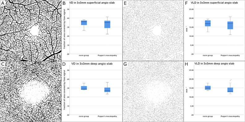
Methods: Twelve patients suffering from poppers maculopathy were included. Health records, optical coherence tomography (OCT), and OCTA data was gathered and compared to a healthy control group (HC). PMP lesion type was determined by manifestation in OCT. OCTA-based evaluation of retinal vascular plexus and choriocapillaris (CC) was executed. Vessel density (VD) and vessel length density (VLD) in superficial and deep capillary plexus (SCP, DCP), as well as flow deficits (FD), within the foveal avascular zone (FAZ) in CC were assessed.

Results: Median age of PMP patients was 40 (min 24; max 64) years, all male. Eleven patients presented with ellipsoid zone-type lesions; one patient showed a vitelliform-type lesion. No qualitative microvascular changes between PMP patients and HC were identified. Quantitative values for VD and VLD of SCP and DCP did not differ in between the two groups. The analysis of FDs in CC showed no deviation from PMP patients to HC.

Conclusions: No vascular anomalies in qualitative and quantitative analysis in OCTA were detected in PMP patients. The constitution of the CC within FAZ of PMP patients does not differ from HC when assessed as FD.

Keywords: Drug toxicity; Multimodal imaging; OCT angiography; Poppers maculopathy; Retinal toxicity.


Conflict of interest statement

The authors declare no competing interests.

Figures

Fig. 1
Fig. 1
Quantification of flow deficits (FD) in the CC: A binarized 6 × 6 mm OCTA slab of the superficial capillary plexus with dotted red ring marking the FAZ; B binarized 6 × 6 OCTA slab of CC with a thickness of 20 μm and an offset of 29 μm below the retinal pigment epithelium (RPE). The red ring lines out the projected FAZ to the CC, which is measured as a .5 mm circle for quantification of FDs in CC = Area1, constituting an area of 0.196 mm2 as a standardized FAZ for all evaluated individuals. The remaining Area2 measure the FDs of perifoveolar ring
Fig. 2
Fig. 2
Quantitative findings of OCTA imaging in poppers maculopathy (PMP) patients (n = 11) compared with age and gender matched healthy controls (n = 11), (AH). Vessel density (VD, as fraction of area covered by flow signal) of superficial and deep capillary plexus (SCP and DCP) was assessed in binarized OCTA images on the respective plexus (A, C) and visualized in box plots (B, D). No significant change was detected between the two groups. Vessel length density (VLD, mm−1) of SCP and DCP measured on skeletonized images (E, G) of the individual plexus did not show any significant difference between the PMP and the healthy group (F, G)
Fig. 3
Fig. 3
Resolution of ellipsoid zone-type PMP lesion over the course of 12 weeks in OCT imaging (AD, F, G). Initial presentation (A, B): note the subfoveal disruption of ellipsoid zone (EZ) and affection of interdigitation zone (IZ). External limiting membrane (ELM) appears continuous but slightly elevated. Similar findings are depicted in 1-week follow-up (C, D). Near infrared en-face imaging (C) demonstrates foveolar hyporeflectivity. After 12 weeks (F, G) sub-foveolar EZ and IZ are assessable as continuous hyperreflective bands but slightly less reflective than the signal of the temporal, nasal continuations of the respective bands. OCTA of superficial capillary plexus of the fovea at weeks 1 and 12 did not show any pathologies (E, H)

References

    1. Davies AJ, Borschmann R, Kelly SP, Ramsey J, Ferris J, Winstock AR. The prevalence of visual symptoms in poppers users: a global survey. BMJ Open Ophthalmol. 2017;1:e000015. doi: 10.1136/bmjophth-2016-000015. - DOI - PMC - PubMed
    1. Davies AJ, Kelly SP, Naylor SG, Bhatt PR, Mathews JP, Sahni J, Haslett R, McKibbin M. Adverse ophthalmic reaction in poppers users: case series of ‘poppers maculopathy’. Eye (Lond) 2012;26:1479–1486. doi: 10.1038/eye.2012.191. - DOI - PMC - PubMed
    1. Pece A, Patelli F, Milani P, Pierro L. Transient visual loss after amyl Isobutyl nitrite abuse. Semin Ophthalmol. 2004;19:105–106. doi: 10.1080/08820530490882292. - DOI - PubMed
    1. Van Bol LB, Kurt RA, Keane PA, Pal B, Sivaprasad S. Clinical phenotypes of poppers maculopathy and their links to visual and anatomic recovery. Ophthalmology. 2017;124:1425–1427. doi: 10.1016/j.ophtha.2017.05.021. - DOI - PubMed
    1. Winstock A.R. BMJea (2019) Global drug survey. https://issuu.com/globaldrugsurvey/docs/gds2019_key_findings_report_may_16_. Accessed 02 Feb 2021