

TBK1 haploinsufficiency results in changes in the K63-ubiquitination profiles in brain and fibroblasts from affected and presymptomatic mutation carriers
- PMID: 34800171
- PMCID: PMC9120096
- DOI: 10.1007/s00415-021-10887-x
TBK1 haploinsufficiency results in changes in the K63-ubiquitination profiles in brain and fibroblasts from affected and presymptomatic mutation carriers
Abstract
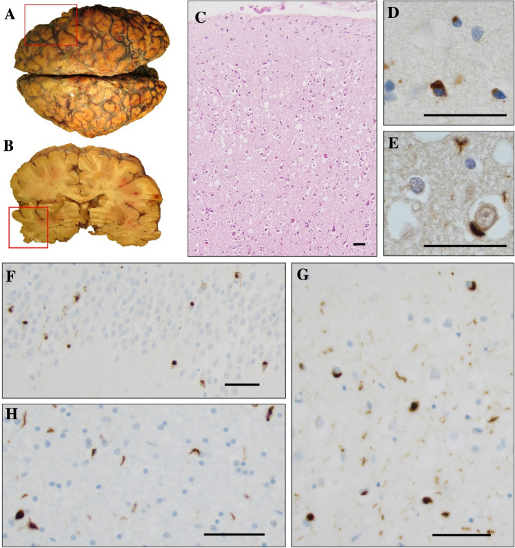
Background: Frontotemporal dementia (FTD) is a neurodegenerative disease, resulting in progressive problems in language and/or behaviour and is often diagnosed before 65 years of age. Ubiquitin positive protein aggregates in the brain are among the key pathologic hallmarks of frontotemporal lobar degeneration (FTLD) postmortem. The TANK-binding kinase 1 gene (TBK1) is on the list of genes that can contribute to the development of FTD as well as the related neurodegenerative disease amyotrophic lateral sclerosis (ALS).
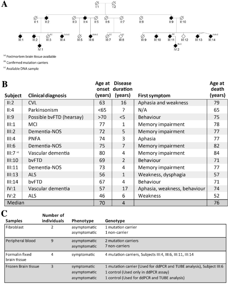
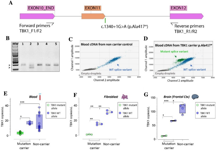
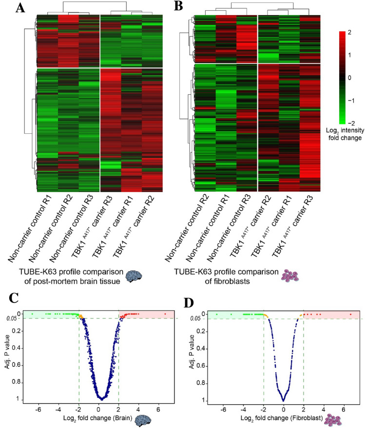
Methods: In this study, using an array of clinical and neuropathological data combined with biochemical and proteomics assays, we analyze the TBK1 splice-mutation (c.1340 + 1G > A) in a Swedish family with a history of FTD and ALS. We also explore the K63 ubiquitination landscape in post-mortem brain tissue and fibroblast cultures.
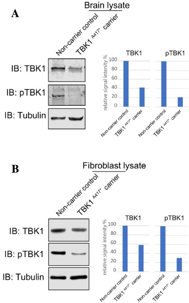
Results: The intronic (c.1340 + 1G > A) mutation in TBK1 results in haploinsufficiency and affects the activity of the protein in symptomatic and pre-symptomatic mutation carriers.
Conclusion: Our results suggest that the mutation leads to a significant reduction of TBK1 activity and induce alterations in K63 ubiquitination profile of the cell already in the presymptomatic stages.
Keywords: ALS; Autophagy; FTD; FTLD; Frontotemporal dementia; Haploinsufficiency; Neurodegeneration; TBK1; Ubiquitination.
© 2021. The Author(s).
Conflict of interest statement
We declare that we have no conflicts of interest to disclose.
Figures
Similar articles
-
TBK1 mutation frequencies in French frontotemporal dementia and amyotrophic lateral sclerosis cohorts.Neurobiol Aging. 2015 Nov;36(11):3116.e5-3116.e8. doi: 10.1016/j.neurobiolaging.2015.08.009. Epub 2015 Aug 14. Neurobiol Aging. 2015. PMID: 26476236
-
A novel TBK1 mutation in a family with diverse frontotemporal dementia spectrum disorders.Cold Spring Harb Mol Case Stud. 2019 Jun 3;5(3):a003913. doi: 10.1101/mcs.a003913. Print 2019 Jun. Cold Spring Harb Mol Case Stud. 2019. PMID: 31160356 Free PMC article.
-
Functional and structural consequences of TBK1 missense variants in frontotemporal lobar degeneration and amyotrophic lateral sclerosis.Neurobiol Dis. 2022 Nov;174:105859. doi: 10.1016/j.nbd.2022.105859. Epub 2022 Sep 13. Neurobiol Dis. 2022. PMID: 36113750 Review.
-
Clinical features of TBK1 carriers compared with C9orf72, GRN and non-mutation carriers in a Belgian cohort.Brain. 2016 Feb;139(Pt 2):452-67. doi: 10.1093/brain/awv358. Epub 2015 Dec 15. Brain. 2016. PMID: 26674655 Free PMC article.
-
Association of Mutations in TBK1 With Sporadic and Familial Amyotrophic Lateral Sclerosis and Frontotemporal Dementia.JAMA Neurol. 2017 Jan 1;74(1):110-113. doi: 10.1001/jamaneurol.2016.3712. JAMA Neurol. 2017. PMID: 27892983 Review.
Cited by
-
Ubiquitin-specific protease 24 promotes EV71 infection by restricting K63-linked polyubiquitination of TBK1.Virol Sin. 2023 Feb;38(1):75-83. doi: 10.1016/j.virs.2022.11.001. Epub 2022 Nov 2. Virol Sin. 2023. PMID: 36334706 Free PMC article.
References
-
- Piguet O, Hornberger M, Mioshi E, Hodges JR (2011) Behavioural-variant frontotemporal dementia: Diagnosis, clinical staging, and management. Lancet Neurol 10: 162–72. Available from: https://pubmed.ncbi.nlm.nih.gov/21147039/ - PubMed
-
- Neary D, Snowden J, Mann D (2005) Frontotemporal dementia. Lancet Neurol 4: 771–780. Available from: https://pubmed.ncbi.nlm.nih.gov/16239184/ - PubMed
-
- Nakano I (2012) Frontotemporal lobar degeneration (FTLD)—changes of its concept and classification based on aggregated proteins. In: Clinical Neurology. Rinsho Shinkeigaku: 1218–20. Available from: https://pubmed.ncbi.nlm.nih.gov/23196569/ - PubMed
-
- Van Der Zee J, Van Broeckhoven C (2014) Dementia in 2013: Frontotemporal lobar degeneration-building on breakthroughs. Nat Rev Neurol 10: 70–72. Available from: https://pubmed.ncbi.nlm.nih.gov/24394289/ - PubMed
MeSH terms
Substances
Grants and funding
LinkOut - more resources
Full Text Sources
Medical
Miscellaneous
