. 2021 Dec 7;33(12):2464-2483.e18.
doi: 10.1016/j.cmet.2021.11.001. Epub 2021 Nov 19.

Quantitative high-confidence human mitochondrial proteome and its dynamics in cellular context

Affiliations

Quantitative high-confidence human mitochondrial proteome and its dynamics in cellular context

Marcel Morgenstern et al. Cell Metab. .

Abstract

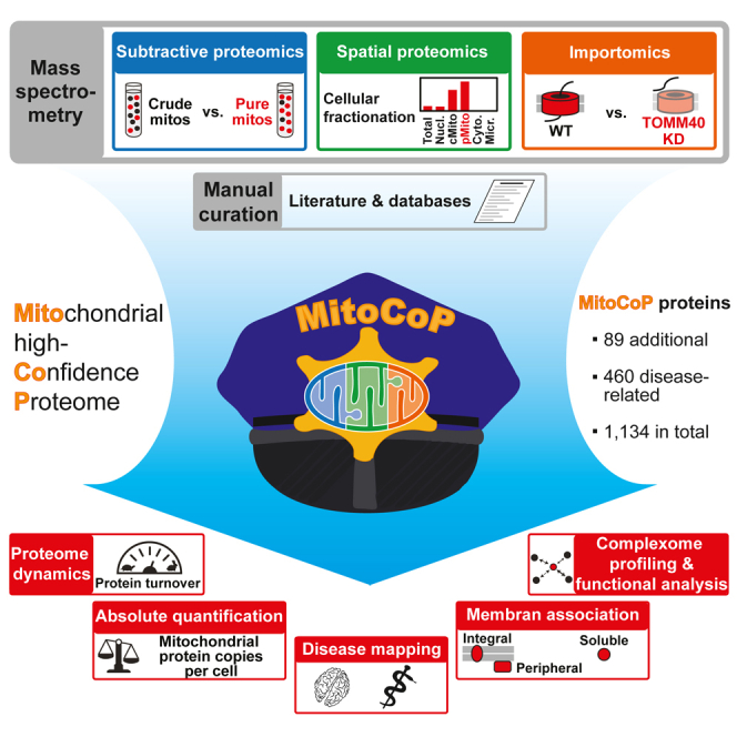
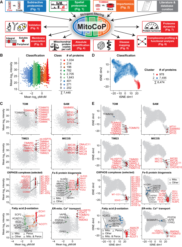
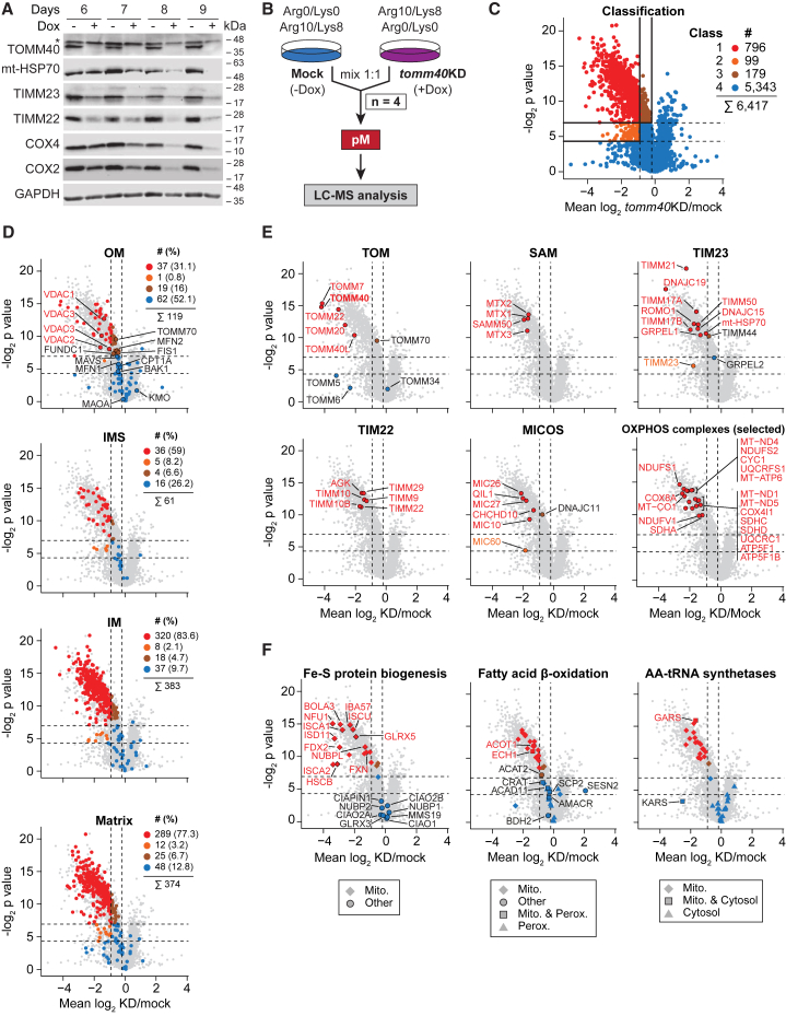
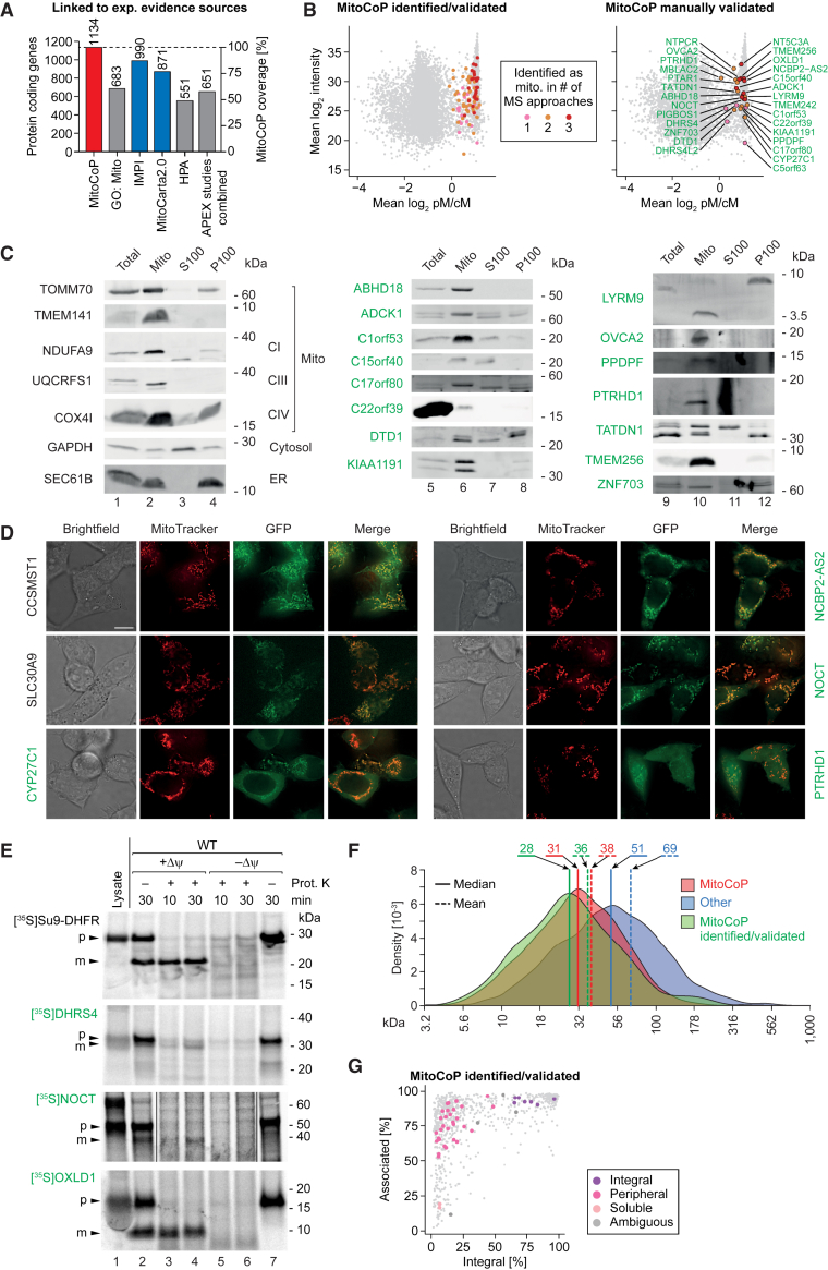
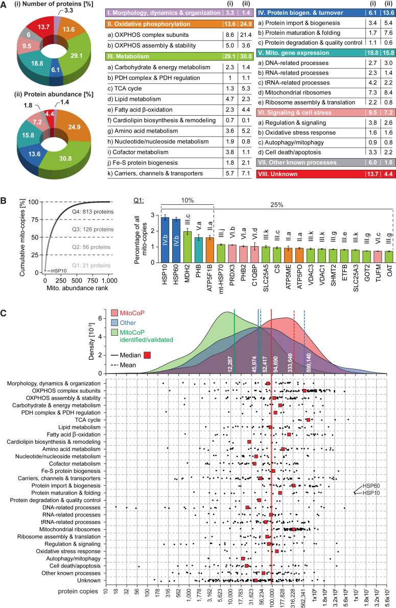
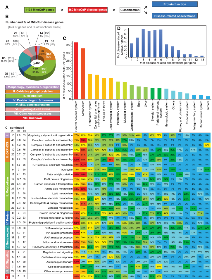
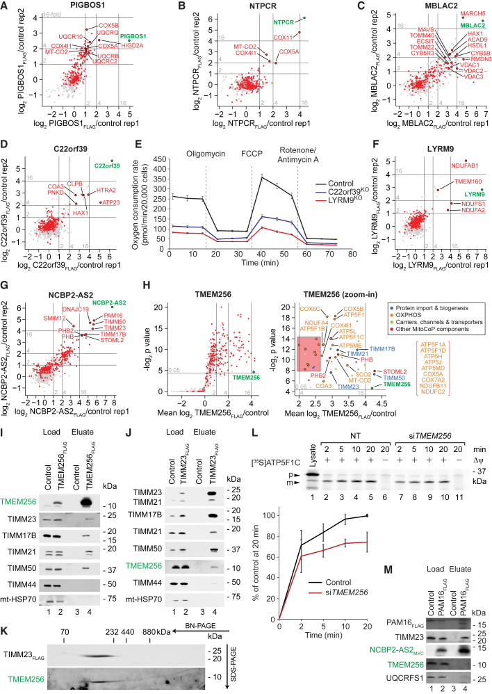
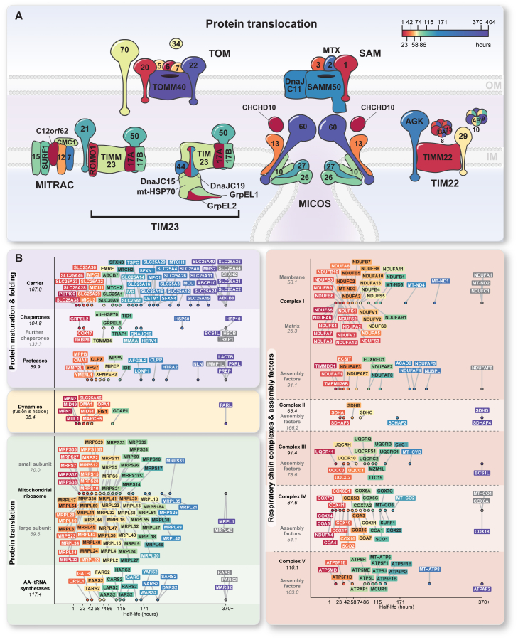
Mitochondria are key organelles for cellular energetics, metabolism, signaling, and quality control and have been linked to various diseases. Different views exist on the composition of the human mitochondrial proteome. We classified >8,000 proteins in mitochondrial preparations of human cells and defined a mitochondrial high-confidence proteome of >1,100 proteins (MitoCoP). We identified interactors of translocases, respiratory chain, and ATP synthase assembly factors. The abundance of MitoCoP proteins covers six orders of magnitude and amounts to 7% of the cellular proteome with the chaperones HSP60-HSP10 being the most abundant mitochondrial proteins. MitoCoP dynamics spans three orders of magnitudes, with half-lives from hours to months, and suggests a rapid regulation of biosynthesis and assembly processes. 460 MitoCoP genes are linked to human diseases with a strong prevalence for the central nervous system and metabolism. MitoCoP will provide a high-confidence resource for placing dynamics, functions, and dysfunctions of mitochondria into the cellular context.

Keywords: Mitochondria; complexome; copy numbers; disease; half-lives; high-confidence proteome; human cells; protein translocation; respiratory chain; smORFs.


Conflict of interest statement

Declaration of interests The authors declare no competing interests.

Figures

None
Graphical abstract
Figure 1
Figure 1
Multifaceted strategy for multidimensional mapping and functional characterization of a high-confidence human mitochondrial proteome (A) Overview of the strategy. Mito., mitochondria; KD, shRNA-mediated knockdown. (B) Ratio-intensity plot of proteins quantified in subtractive proteomics experiments of crude and gradient-purified mitochondria (n ≥ 3/4 biological replicates). Numbers of proteins indicated include isoforms. cM/pM, crude/pure mitochondria. (C) Same as in (B) highlighting selected sets of proteins. Fe-S, iron-sulfur; Perox., peroxisomal; ER, endoplasmic reticulum. (D) Two-dimensional map of subcellular fractionation data generated as shown in Figure S1B. Cluster reflect the distribution of mitochondrial and non-mitochondrial test sets (see Figure S2B). dim, dimension. (E) tSNE plots highlighting selected sets of proteins. dim, dimension. See also Figures S1 and S2 and Table S2.
Figure 2
Figure 2
Charting human mitochondrial proteins by importomics (A) Steady-state levels of mitochondrial proteins in whole cell lysates of doxycycline (Dox)-induced (+) and mock-treated (−) tomm40-shRNA cells. GAPDH, loading control; , non-specific band. (B) Outline of the importomics approach. pM, pure mitochondria; KD, shRNA-mediated knockdown. (C) Volcano plot of proteins quantified by importomics following shRNA-mediated knockdown of tomm40 (n ≥ 2/4 biological replicates). Classes were defined based on the distribution of mitochondrial and non-mitochondrial test sets (see Figures S3D–S3G). Numbers of proteins indicated include isoforms. Horizontal lines mark p values (two-sided Student's t test) of 0.05 (bottom) and 0.0081 (top; Benjamini-Hochberg-corrected); vertical lines mark tomm40KD/mock ratios of 0.53 (left) and 0.87 (right). (D–F) Same as in (C) highlighting proteins of distinct submitochondrial localizations (D), of different mitochondrial protein complexes (E), and of selected functional categories (F). OM/IM, outer/inner mitochondrial membrane; IMS, intermembrane space; AA, aminoacyl. See also Figure S3 and Tables S2 and S3.
Figure 3
Figure 3
The human mitochondrial high-confidence proteome “MitoCoP” (A) Number and coverage of protein-coding genes present in MitoCoP versus major mitochondrial protein repositories/datasets based on entries with experimental evidence for mitochondrial localization. APEX, in situ proximity labeling studies of submitochondrial proteomes (Hung et al., 2014, 2017; Rhee et al., 2013); GO: Mito, GO-CC term “mitochondrion;” IMPI, integrated mitochondrial protein index; HPA, human protein atlas (Thul et al., 2017). (B) Ratio-intensity plots highlighting MitoCoP identified/validated proteins in the subtractive proteomics dataset. (C) Subcellular fractions of HEK293T cells were analyzed by western blotting using antibodies directed against the indicated marker (black) and MitoCoP identified/validated proteins (green). CI, CIII, and CIV, respiratory complexes I, III, and IV; Mito, mitochondrial fraction; S100, cytosolic fraction; P100, microsomal fraction. (D) HEK293T cells were transfected with vectors carrying GFP-tagged proteins and MitoTracker Red was added to visualize the mitochondrial network. Live cell images of the GFP signal (green) were recorded with the mitochondrial signal (red) and merged. Scale bar, 10 μm. (E) In organello import of radiolabeled precursor proteins into HEK293T mitochondria in the presence or absence of a membrane potential (Δψ), followed by proteinase K (Prot. K) treatment. In case of NOCT, the + Prot. K gel lanes were exposed ∼1.5-times longer than the other gel lanes. p, precursor protein; m, mature protein. Lysate, in vitro synthesized radiolabeled precursor protein. (F) Molecular mass distribution of MitoCoP proteins, the remaining cellular proteome (other; excluding MitoCoP proteins), and MitoCoP identified/validated proteins. (G) Membrane association of MitoCoP proteins (see also Figures S4D–S4F). The mitochondrial localization of proteins labeled in green has been confirmed experimentally in this study by biochemical assays, fluorescence microscopy, and/or q-AP-MS experiments (Figure 6). See also Figure S4 and Tables S1 and S3.
Figure 4
Figure 4
Functional and absolute quantitative portrait of MitoCoP (A) Functional classification of MitoCoP comprising 1,134 proteins. Protein abundance (ii) reflects mito-copy numbers per cell. PDH, pyruvate dehydrogenase; TCA, tricarboxylic acid; biogen., biogenesis. (B) Cumulative mito-copy number plot (left) with quantitative and functional information about abundant MitoCoP proteins accounting for 25% (i.e., Q1) of total mito-copy numbers per cell (right). Error bars, SEM for n = 3 and range for n = 2. (C) Copy number distributions of MitoCoP proteins, the remaining cellular proteome (other; excluding MitoCoP proteins) and MitoCoP identified/validated proteins (top) and of individual MitoCoP constituents grouped according to functional classes as defined in (A) (bottom). See also Figures S5A–S5E and Tables S1 and S4.
Figure 5
Figure 5
MitoCoP disease gene classification (A) For disease mapping all MitoCoP genes were screened for disease association and classified according to protein function and disease-related observations. (B) Functional classification of the MitoCoP disease genes as in Figure 4A. (C) Top: Number of MitoCoP disease genes associated with different disease-related observations. Bottom: Heatmap indicating the occurrence of specific observations for MitoCoP disease genes related to their functional classification. (#), number and percentage (i) of mitochondrial disease genes for the functional sub-/class; (ii), percentage of the disease genes related to all genes of the functional sub-/class. (D) Number of MitoCoP genes associated with one or multiple disease-related observations per gene. See also Figures S5F and S5G and Table S1.
Figure 6
Figure 6
Functional MitoCoP interaction networks (A–D, F, and G) Interaction networks of selected MitoCoP proteins analyzed by q-AP-MS (n = 2). (E) Oxygen consumption rate of control, C22orf39KO, and LYRM9KO HEK293T cells after the indicated treatments. Error bars, SEM (n = 12). FCCP, carbonyl cyanide 4-(trifluoromethoxy)phenylhydrazone. (H) q-AP-MS analysis of TMEM256FLAG interacting proteins (n = 4). P values were determined using a one-sided Student's t test. (I) Cells transiently expressing TMEM256FLAG were lysed with digitonin and subjected to FLAG-immunoprecipitation and eluates were analyzed by SDS-PAGE. (Load = 1%, Eluate = 100%). (J and K) Mitochondria isolated from TIM23FLAG expressing cells were subjected to FLAG-immunoprecipitation. Bound complexes were eluted natively and analyzed by SDS-PAGE (J) (Load = 1%, Eluate = 100%) or 2D-BN/SDS-PAGE (K) (Eluate = 100%). (L) Mitochondria from TMEM256-depleted cells were isolated prior import of [35S]ATPC1 precursor followed by proteinase K treatment, SDS-PAGE, and digital autoradiography. Error bars, SEM (n = 3); p, precursor; m, mature; Lysate, synthesized precursor. (M) FLAG-immunoprecipitation eluates of NCBP2-AS2MYC and PAM16FLAG expressing cells lysed with digitonin were analyzed by SDS-PAGE. (Load = 2%, Eluate = 100%). See also Figure S6J and Table S5.
Figure 7
Figure 7
Protein half-life map of the human mitochondrial organizing network (A) Schematic illustration of the two membrane-spanning mitochondrial protein import systems showing half-lives for individual components of each complex. CHCHD10/MIC14 is possibly linked to MICOS. IM/OM, inner/outer mitochondrial membrane; MITRAC, mitochondrial translation regulation assembly intermediate of cytochrome c oxidase. (B) Protein half-life landscape of the central mitochondrial complexome grouped according to function. Numbers in italics indicate the median half-life of the respective protein group. See also Figure S7 and Table S6.

References

    1. Anand R., Strecker V., Urbach J., Wittig I., Reichert A.S. Mic13 Is Essential for Formation of Crista Junctions in Mammalian Cells. PLoS One. 2016;11:e0160258. - PMC - PubMed
    1. Angerer H. Eukaryotic LYR Proteins Interact with Mitochondrial Protein Complexes. Biology (Basel) 2015;4:133–150. - PMC - PubMed
    1. Anton F., Dittmar G., Langer T., Escobar-Henriques M. Two deubiquitylases act on mitofusin and regulate mitochondrial fusion along independent pathways. Mol. Cell. 2013;49:487–498. - PubMed
    1. Araiso Y., Tsutsumi A., Qiu J., Imai K., Shiota T., Song J., Lindau C., Wenz L.-S., Sakaue H., Yunoki K., et al. Structure of the mitochondrial import gate reveals distinct preprotein paths. Nature. 2019;575:395–401. - PubMed
    1. Ashburner M., Ball C.A., Blake J.A., Botstein D., Butler H., Cherry J.M., Davis A.P., Dolinski K., Dwight S.S., Eppig J.T., et al. The Gene Ontology Consortium Gene ontology: tool for the unification of biology. Nat. Genet. 2000;25:25–29. - PMC - PubMed

Publication types

MeSH terms