. 2021 Dec;297(6):101426.
doi: 10.1016/j.jbc.2021.101426. Epub 2021 Nov 17.

FBXO7 triggers caspase 8-mediated proteolysis of the transcription factor FOXO4 and exacerbates neuronal cytotoxicity

Affiliations

FBXO7 triggers caspase 8-mediated proteolysis of the transcription factor FOXO4 and exacerbates neuronal cytotoxicity

Su Hyoun Lee et al. J Biol Chem. 2021 Dec.

Abstract

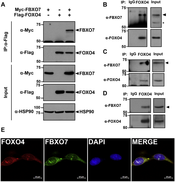
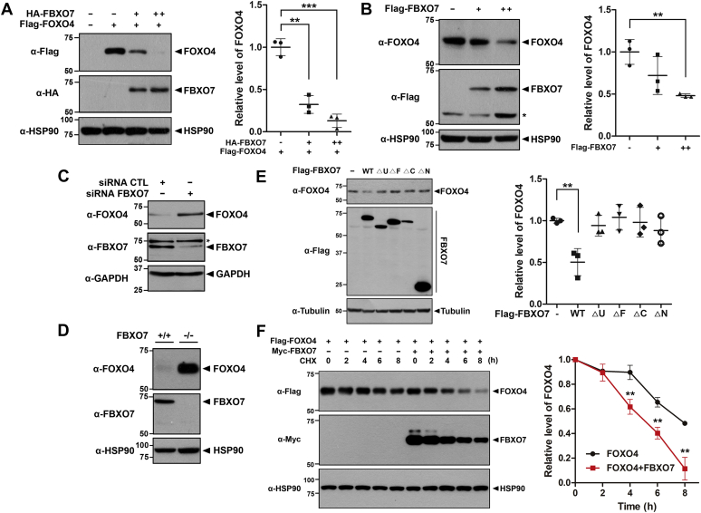
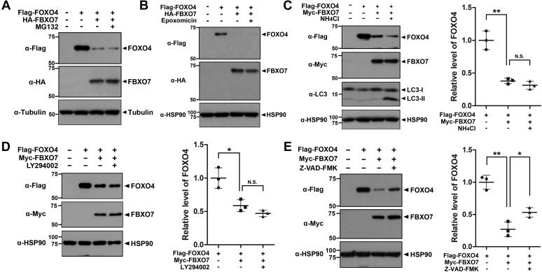
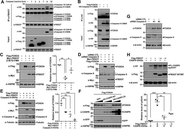
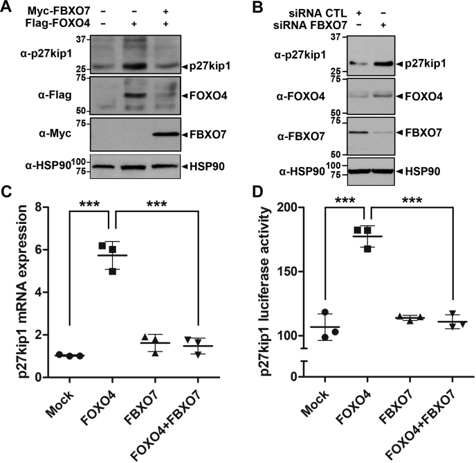
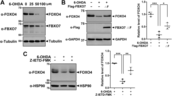
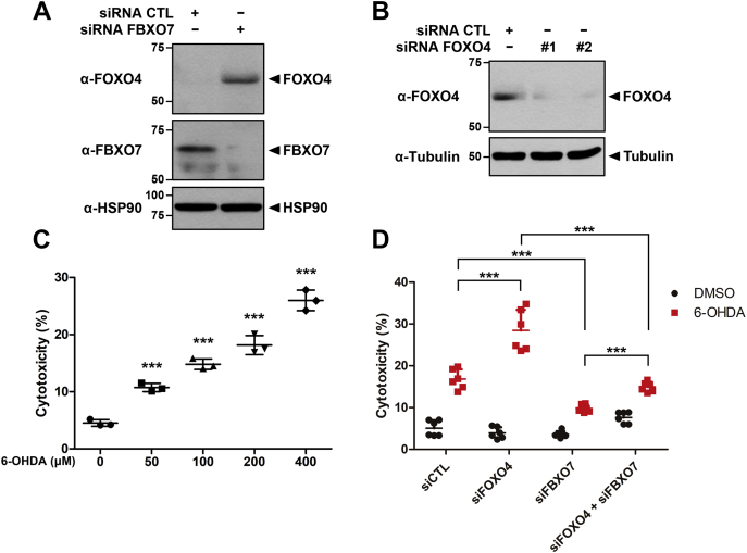
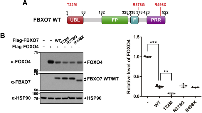
Parkinson's disease (PD) is characterized by the progressive loss of midbrain dopamine neurons in the substantia nigra. Mutations in the F-box only protein 7 gene (Fbxo7) have been reported to cause an autosomal recessive form of early-onset familial PD. FBXO7 is a part of the SKP1-Cullin1-F-box (SCF) E3 ubiquitin ligase complex, which mediates ubiquitination of numerous substrates. FBXO7 also regulates mitophagy, cell growth, and proteasome activity. A member of the FOXO family, the transcription factor FOXO4, is also known to modulate several cellular responses, including cell cycle progression and apoptosis; however, the relationship between FBXO7 and FOXO4 has not been investigated. In this study, we determined that FBXO7 binds to FOXO4 and negatively regulates intracellular FOXO4 levels. Interestingly, we also found that FBXO7-mediated degradation of FOXO4 did not occur through either of two major proteolysis systems, the ubiquitin-proteasome system or the lysosome-autophagy pathway, although it was blocked by a caspase 8-specific inhibitor and caspase 8-knockdown. Moreover, intracellular FOXO4 levels were greatly reduced in dopaminergic MN9D cells following treatment with neurotoxic 6-hydroxydopamine (6-OHDA), which was produced upon FBXO7-mediated and caspase 8-mediated proteolysis. Taken together, these results suggest that FOXO4 is negatively regulated in FBXO7-linked PD through caspase 8 activation, suppressing the cytoprotective effect of FOXO4 during 6-OHDA-induced neuronal cell death.

Keywords: 6-OHDA; FBXO7; FOXO4; Parkinson’s disease; caspase 8; neuronal cell death.


Conflict of interest statement

Conflict of interest The authors declare that they have no conflict of interest with the content of this article.

Figures

Figure 1
Figure 1
FBXO7 interacts with FOXO4.A, HEK293 cell were transfected for 24 h with plasmids encoding Myc-FBXO7 and/or FLAG-FOXO4. Cell lysates were immunoprecipitated using an anti-FLAG antibody and the precipitates immunoblotted with the indicated antibodies. HSP90 served as a loading control. B, HEK293 cell-lysates were immunoprecipitated using either an anti-FOXO4 antibody or preimmune IgG (control), followed by immunoblotting with the indicated antibodies. C, MN9D cell lysates were immunoprecipitated using an anti-FOXO4 IgG, followed by immunoblotting with the indicated antibodies. D, mouse brain lysates were immunoprecipitated using anti-FOXO4 IgG, followed by immunoblotting with the indicated antibodies. E, SH-SY5Y cells were fixed, permeabilized, and immunostained. Representative confocal images of cells immunostained for endogenous FOXO4 (red) and endogenous FBXO7 (green) are shown. Nuclei were counterstained with DAPI (blue). Scale bars denote 10 μm.
Figure 2
Figure 2
FBXO7 decreases FOXO4 protein stability.A, HEK293 cells were transfected for 24 h with a plasmid encoding FLAG-FOXO4, either alone or in combination with increasing amounts of a plasmid encoding HA-FBXO7. Transfected cell lysates were immunoblotted and screened with anti-FLAG or anti-HA antibodies. Relative levels of FOXO4 were quantified and the results presented as the mean ± SD of three independent experiments (∗∗∗p ≤ 0.0001; ∗∗p ≤ 0.001). B, HEK293 cells were transfected for 24 h with increasing amounts of plasmid encoding FLAG-FBXO7. Transfected cell lysates were immunoblotted and screened with anti-FOXO4 or anti-FLAG antibodies. Relative levels of FOXO4 were quantified and results presented as the mean ± SD of three independent experiments (∗∗p ≤ 0.001). C, HEK293 cells were transfected for 48 h with siRNA-control (CTL) or siRNA-FBXO7. Transfected cell lysates were immunoblotted and screened with anti-FOXO4 or anti-FBXO7 antibodies. D, Immunoblotting analysis of lysates of FBXO7+/+ and FBXO7−/− HAP1 cells was performed using anti-FOXO4 or anti-FBXO7 antibodies. E, HEK293 cells were transfected for 24 h with a plasmid encoding FLAG-tagged wild-type FBXO7 or its deletion mutants. Transfected cell lysates were immunoblotted and screened with anti-FOXO4 or anti-Flag antibodies. Relative levels of FOXO4 were quantified and the results presented as the mean ± SD of three independent experiments (∗∗p ≤ 0.001). F, HEK293 cells were transfected for 24 h with plasmids encoding FLAG-FOXO4 and/or Myc-FBXO7. Cells were treated for the indicated times with 25 μg/ml cycloheximide, and cell lysates were immunoblotted with the indicated antibodies. Relative levels of FOXO4 were quantified and the results presented as the mean ± SD of three independent experiments (∗∗p ≤ 0.001). Hsp90, GAPDH, and Tubulin served as loading controls.
Figure 3
Figure 3
FBXO7 decreases FOXO4 stability through caspase activation.A and B, HEK293 cells were transfected for 24 h with plasmids encoding FLAG-FOXO4 and/or HA-FBXO7. Cells were then treated for an additional 6 h with vehicle (−), 10 μM MG132 (A), or 1 μM epoxomicin (B). C, HEK293 cells were transfected for 24 h with plasmids encoding FLAG-FOXO4 and/or Myc-FBXO7. Cells were then treated for additional 6 h with vehicle (−) or 25 mM NH4Cl. Relative levels of FOXO4 were quantified and the results presented as the mean ± SD of three independent experiments (∗∗p ≤ 0.001; N.S., not significant). D, HEK293 cells were transfected for 24 h with plasmids encoding FLAG-FOXO4 and/or Myc-FBXO7. Cells were then treated for additional 4 h with vehicle (−) or 50 μM LY294002. Relative levels of FOXO4 were quantified and the results presented as the mean ± SD of three independent experiments (∗p ≤ 0.05; N.S., not significant). E, HEK293 cells were transfected for 24 h with plasmids encoding FLAG-FOXO4 and/or Myc-FBXO7. The cells were then treated for additional 6 h with vehicle (−) or 100 μM Z-VAD-FMK. H, relative levels of FOXO4 were quantified and the results presented as the mean ± SD of three independent experiments (∗∗p ≤ 0.001; ∗p ≤ 0.05). Tubulin and Hsp90 served as a loading control.
Figure 4
Figure 4
FBXO7 decreases FOXO4 stability through a caspase 8 activation.A, HEK293 cells were transfected for 24 h with the plasmids encoding FLAG-caspase 1-C284A, FLAG-caspase 2-C303A, FLAG-caspase 3-C163A, FLAG-caspase 7-C186A, HA-caspase 8-C360S, or HA-caspase 10-C401A. Cell lysates were immunoprecipitated using anti-FBXO7 antibody followed by immunoblotting with the indicated antibodies. B, HEK293 cells were transfected for 24 h with plasmids encoding HA-caspase 8-C360S and/or FLAG-FOXO4. Cell lysates were immunoprecipitated with anti-HA antibody. The precipitates were immunoblotted screened using the indicated antibodies. C, HEK293 cells were transfected for 24 h with plasmids encoding FLAG-FOXO4 and/or Myc-FBXO7. The cells were then treated for additional 3 h with vehicle (−) or Z-IETD-FMK (20 μM). Relative levels of FOXO4 were quantified and the results presented as the mean ± SD of three independent experiments (∗∗p ≤ 0.001; ∗, p ≤ 0.05). D, HEK293 cells were transfected for 48 h with siRNA-control (CTL), siRNA-caspase 8, a plasmid encoding FLAG-FOXO4, or a plasmid encoding Myc-FBXO7, either alone or in combination. Cell lysates were immunoblotted and screened using the indicated antibodies. E, HEK293 cells were transfected for 24 h with plasmids encoding FLAG-FOXO4, Myc-FBXO7, or caspase 8-V5/His, either alone or in combination. Cell lysates were immunoblotted and screened using the indicated antibodies. Relative levels of FOXO4 were quantified and the results presented as the mean ± SD of three independent experiments (∗∗∗p ≤ 0.0001; ∗∗p ≤ 0.001). F, HEK293 cells were transfected for 24 h with a plasmid encoding FLAG-FOXO4, either alone or in combination with increasing amounts of a plasmid encoding HA-tagged wild-type caspase 8 (CASP8) or its C360S mutant. Transfected cell lysates were immunoblotted and screened using the indicated antibodies. G, HEK293 cells were transfected for 48 h with control siRNA (CTL), caspase 8-siRNA #1 or #2. Transfected cell lysates were immunoblotted and screened with anti-FOXO4 or anti-caspase 8 antibodies. H, HEK293 cells were transfected for 24 h with a plasmid encoding caspase 8-V5/His, Flag-FBXO7-WT, Flag-FBXO7-ΔU, or Flag-FBXO7-ΔF alone or in combination. Cell lysates were immunoblotted and screened using the indicated antibodies. Relative levels of p43/41 were quantified and the results presented as the mean ± SD of three independent experiments (∗∗∗, p ≤ 0.0001; ∗∗, p ≤ 0.001). Hsp90, β-actin, and Tubulin served as loading controls.
Figure 5
Figure 5
FBXO7 decreases the transcription activity of FOXO4.A, HEK293 cells were transfected for 24 h with a plasmid encoding FLAG-FOXO4, either alone or in combination with a plasmid encoding Myc-FBXO7. Transfected cell lysates were immunoblotted and screened using anti-p27kip1, anti-FLAG, or anti-Myc antibody. B, HEK293 cells were transfected for 48 h with siRNA-control (CTL), or siRNA-FBXO7, and the cell lysates were immunoblotted and screened using anti-p27kip1, anti-FOXO4, or anti-FBXO7 antibody. Hsp90 served as a loading control. C, HEK293 cells were transfected for 24 h with plasmids encoding FLAG-FOXO4 and/or Myc-FBXO7. Total RNA was extracted and reverse transcribed. Levels of p27kip1 mRNA were assessed using real-time PCR. Data are presented as the mean ± SD of three independent experiments (∗∗∗p ≤ 0.0001). D, MCF7 cells were cotransfected with the pGL3-p27kip1-Luc plasmid and the indicated plasmids. Following the incubation for 24 h, firefly luciferase activity was measured and normalized to the Renilla luciferase activity. Data are presented as the mean ± SD of three independent experiments (∗∗∗p ≤ 0.0001).
Figure 6
Figure 6
Treatment with 6-OHDA reduces FOXO4 levels via FBXO7-mediated proteolysis.A, MN9D cells were treated for 6 h with vehicle (−) or 6-OHDA. Cell lysates were then immunoblotted with anti-FOXO4 or anti-FBXO7 antibodies. B, MN9D cells were transfected for 24 h with a plasmid encoding FLAG-FBXO7-ΔF mutant and then treated for additional 6 h with vehicle (−) or 6-OHDA (50 μM). Cell lysates were immunoblotted and screened using anti-FOXO4 or anti-FLAG antibodies. Relative levels of FOXO4 were quantified and the results presented as the mean ± SD of three independent experiments (∗∗∗p ≤ 0.0001; ∗p ≤ 0.05). C, MN9D cells were left untreated or treated for 3 h with 6-OHDA (50 μM), either alone or in combination with Z-IETD-FMK (20 μM). Cell lysates were immunoblotted and screened using anti-FOXO4 antibody. Relative levels of FOXO4 were quantified and the results presented as the mean ± SD of three independent experiments (∗∗∗p ≤ 0.0001; ∗∗p ≤ 0.001). Tubulin, GAPDH, and Hsp90 served as loading controls.
Figure 7
Figure 7
FBXO7 reduces cytoprotective effects of FOXO4 through 6-OHDA-induced cell death.A, MN9D cells were transfected for 48 h with control siRNA (CTL) or FBXO7-siRNA, and the cell lysates were immunoblotted and screened using anti-FOXO4 or anti-FBXO7 antibodies. B, MN9D cells were transfected for 48 h with control siRNA (CTL), FOXO4-siRNA #1, or FOXO4-siRNA #2. Cell lysates were immunoblotted and screened using the indicated antibodies. Hsp90 and Tubulin served as loading controls. C, MN9D cells were treated for 4 h with DMSO (vehicle) or the indicated concentrations of 6-OHDA. Cell toxicity was measured using LDH assays. Data are presented as the mean ± SD of three independent experiments (∗∗∗p ≤ 0.0001). D, MN9D cells were transfected for 48 h with siRNA-control (CTL), siRNA-FOXO4, or siRNA-FBXO7 and then treated for additional 4 h with DMSO or 6-OHDA (100 μM). Data are presented as the mean ± SD of six independent experiments (∗∗∗p ≤ 0.0001).
Figure 8
Figure 8
The PD-linked FBXO7-T22M mutant reduces FOXO4 stability much greater than wild-type FBXO7.A, Schematic of PD-linked mutations in FBXO7. B, HEK293 cells were transfected for 24 h with plasmids encoding FLAG-FOXO4, wild-type FLAG-FBXO7, mutant FLAG-FBXO7-T22M, mutant FLAG-FBXO7-R378G, or mutant FLAG-FBXO7-R498X, either alone or in combination. Cell lysates were immunoblotted and screened using the indicated antibodies. Relative levels of FOXO4 were quantified and the results presented as the mean ± SD of three independent experiments (∗∗∗p ≤ 0.0001; ∗∗p ≤ 0.001). Hsp90 served as a loading control.

References

    1. de Lau L.M.L., Breteler M.M.B. Epidemiology of Parkinson's disease. Lancet Neurol. 2016;5:525–535. - PubMed
    1. Wakabayashi K., Tanji K., Mori F., Takahashi H. The Lewy body in Parkinson’s disease: Molecules implicated in the formation and degradation of α-synuclein aggregates. Neuropathology. 2007;27:494–506. - PubMed
    1. Antony P.M., Diederich N.J., Kruger R., Balling R. The hallmarks of Parkinson’s disease. FEBS J. 2013;280:5981–5993. - PubMed
    1. Zhou Z.D., Lee J.C.T., Tan E.K. Pathophysiological mechanisms linking F-box only protein 7 (FBXO7) and Parkinson's disease (PD) Mutat. Res. 2018;778:72–78. - PubMed
    1. Kipreos E.T., Pagano M. The F-box protein family. Genome Biol. 2000;1 reviews3002.1-3002.7. - PMC - PubMed

Publication types