. 2021 Nov 20;13(1):137.
doi: 10.1186/s13098-021-00753-1.

CircSMAD4 alleviates high glucose-induced inflammation, extracellular matrix deposition and apoptosis in mouse glomerulus mesangial cells by relieving miR-377-3p-mediated BMP7 inhibition

Affiliations

CircSMAD4 alleviates high glucose-induced inflammation, extracellular matrix deposition and apoptosis in mouse glomerulus mesangial cells by relieving miR-377-3p-mediated BMP7 inhibition

Rina Wu et al. Diabetol Metab Syndr. .

Abstract

Background: Diabetic nephropathy (DN) is a common complication of diabetes mellitus. Accumulating studies suggest that the deregulation of circular RNA (circRNA) is involved in DN pathogenesis. This study aimed to investigate the role of circSMAD4 in DN models.

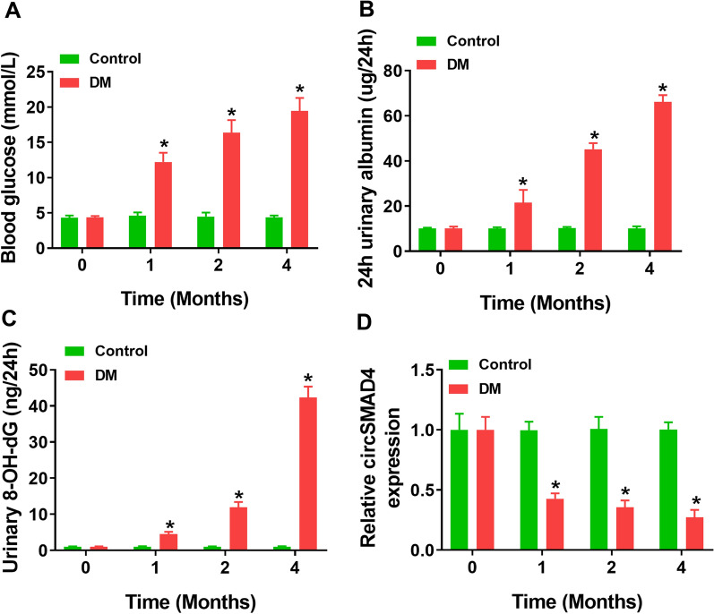
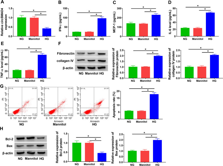
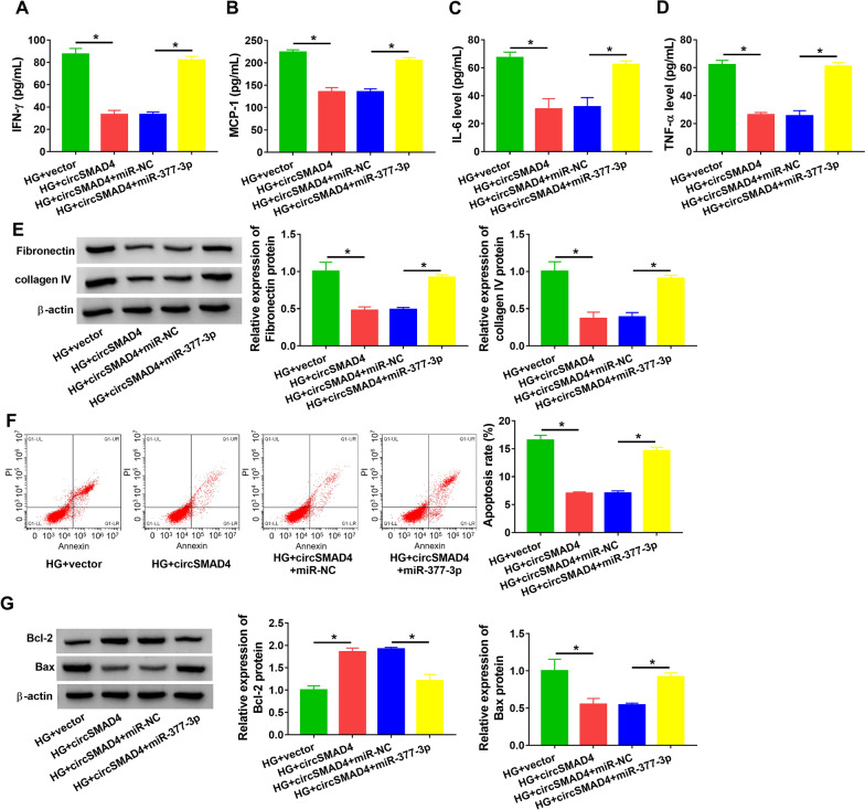
Methods: Mice were treated with streptozotocin to establish DN models in vivo. Mouse glomerulus mesangial cells (SV40-MES13) were treated with high glucose to establish DN models in vitro. The expression of circSMAD4, miR-377-3p and bone morphogenetic protein 7 (BMP7) mRNA was measured by quantitative real-time PCR (qPCR). The releases of inflammatory factors were examined by ELISA. The protein levels of fibrosis-related markers, apoptosis-related markers and BMP7 were checked by western blot. Cell apoptosis was monitored by flow cytometry assay. The predicted relationship between miR-377-3p and circSMAD4 or BMP7 was validated by dual-luciferase reporter assay or pull-down assay.

Results: CircSMAD4 was poorly expressed in DN mice and HG-treated SV40-MES13 cells. HG induced SV40-MES13 cell inflammation, extracellular matrix (ECM) deposition and apoptosis. CircSMAD4 overexpression alleviated, while circSMAD4 knockdown aggravated HG-induced SV40-MES13 cell injuries. MiR-377-3p was targeted by circSMAD4, and miR-377-3p enrichment partly reversed the effects of circSMAD4 overexpression. BMP7 was a target of miR-377-3p, and circSMAD4 regulated BMP7 expression by targeting miR-377-3p. MiR-377-3p overexpression aggravated HG-induced injuries by suppressing BMP7.

Conclusion: CircSMAD4 alleviates HG-induced SV40-MES13 cell inflammation, ECM deposition and apoptosis by relieving miR-377-3p-mediated inhibition on BMP7 in DN progression.

Keywords: BMP7; Diabetic nephropathy; circSMAD4; miR-377-3p.


Conflict of interest statement

The authors declare that they have no conflicts of interest.

Figures

Fig. 1
Fig. 1
CircSMAD4 expression was decreased in DN models. A Blood glucose was measured in mice. B and C Urinary albumin and urinary 8-OH-dG were measured by ELISA. D The expression of circSMAD4 in kidney tissues was detected by qPCR. *P < 0.05
Fig. 2
Fig. 2
HG induced SV40-MES13 cell inflammation, ECM deposition and cell apoptosis. SV40-MES13 cells were treated with HG, NG or mannitol. A The expression of circSMAD4 was detected by qPCR. BE The releases of IFN-γ, MCP-1, IL-6 and TNF-α in medium were determined by ELISA. F The protein levels of fibronectin and collagen IV were measured by western blot. G Cell apoptosis was assessed by flow cytometry assay. H The protein levels of Bcl-2 and Bax were measured by western blot. *P < 0.05
Fig. 3
Fig. 3
CircSMAD4 overexpression alleviated HG-induced SV40-MES13 cell inflammation, ECM deposition and cell apoptosis. A The efficiency of circSMAD4 overexpression or knockdown was checked by qPCR. HG-treated SV40-MES13 cells were transfected with circSMAD4 or sh-circSMAD4. Then, B the expression of circSMAD4 was detected by qPCR. CF The releases of IFN-γ, MCP-1, IL-6 and TNF-α in medium were determined by ELISA. G The protein levels of fibronectin and collagen IV were measured by western blot. H Cell apoptosis was assessed by flow cytometry assay. I The protein levels of Bcl-2 and Bax were measured by western blot. *P < 0.05
Fig. 4
Fig. 4
MiR-377-3p was a target of circSMAD4. A The binding relationship between circSMAD4 and miR-377-3p was predicted by starbase. B The efficiency of miR-377-3p overexpression or inhibition was checked by qPCR. C and D The binding relationship between circSMAD4 and miR-377-3p was verified by dual-luciferase reporter assay and pull-down assay. E The expression of miR-377-3p in SV40-MES13 cells was treated with HG, NG or mannitol was detected by qPCR. F MiR-377-3p expression in HG-treated SV40-MES13 cells with circSMAD4 absence was checked by qPCR. *P < 0.05
Fig. 5
Fig. 5
CircSMAD4 overexpression alleviated HG-induced SV40-MES13 cell inflammation, ECM deposition and cell apoptosis by impairing miR-377-3p expression. AG SV40-MES13 cells treated with HG were transfected with circSMAD4 or circSMAD4 + miR-377-3p. A–D The releases of IFN-γ, MCP-1, IL-6 and TNF-α in medium were determined by ELISA. E The protein levels of fibronectin and collagen IV were measured by western blot. F Cell apoptosis was assessed by flow cytometry assay. G The protein levels of Bcl-2 and Bax were measured by western blot. *P < 0.05
Fig. 6
Fig. 6
CircSMAD4 regulated BMP7 expression by targeting miR-377-3p. A MiR-377-3p binding to BMP7 3′UTR was predicted by DIANA tools. B The interaction between miR-377-3p and BMP7 was predicted by dual-luciferase reporter assay. C The expression of BMP7 protein in SV40-MES13 cells treated with HG, NG or mannitol was detected by western blot. D The expression of BMP7 protein in SV40-MES13 cells transfected with miR-377-3p or anti-miR-377-3p was measured by western blot. E The expression of BMP7 protein in SV40-MES13 cells transfected with sh-circSMAD4 or sh-circSMAD4 + anti-miR-377-3p was detected by western blot. *P < 0.05
Fig. 7
Fig. 7
MiR-377-3p overexpression aggravated HG-induced SV40-MES13 cell inflammation, ECM deposition and cell apoptosis by suppressing BMP7. A The efficiency of BMP7 overexpression was tested by western blot. BI HG-treated SV40-MES13 cells were transfected with miR-377-3p or miR-377-3p + BMP7. B The expression of BMP7 protein was detected by western blot. CF The releases of IFN-γ, MCP-1, IL-6 and TNF-α in medium were determined by ELISA. G The protein levels of fibronectin and collagen IV were measured by western blot. H Cell apoptosis was assessed by flow cytometry assay. I The protein levels of Bcl-2 and Bax were measured by western blot. *P < 0.05

Similar articles

Cited by

References

    1. Khan NU, et al. Insights into predicting diabetic nephropathy using urinary biomarkers. Biochim Biophys Acta Proteins Proteom. 2020;1868(10):140475. doi: 10.1016/j.bbapap.2020.140475. - DOI - PubMed
    1. Xiong Y, Zhou L. The signaling of cellular senescence in diabetic nephropathy. Oxid Med Cell Longev. 2019;2019:7495629. - PMC - PubMed
    1. Gheith O, et al. Diabetic kidney disease: world wide difference of prevalence and risk factors. J Nephropharmacol. 2016;5(1):49–56. - PMC - PubMed
    1. Lu Z, Liu N, Wang F. Epigenetic regulations in diabetic nephropathy. J Diabetes Res. 2017;2017:7805058. - PMC - PubMed
    1. Umanath K, Lewis JB. Update on diabetic nephropathy: core curriculum 2018. Am J Kidney Dis. 2018;71(6):884–895. doi: 10.1053/j.ajkd.2017.10.026. - DOI - PubMed

LinkOut - more resources