

doi: 10.1016/S2213-8587(21)00291-6.
Epub 2021 Nov 18.
COVID-19 targets human adrenal glands
Affiliations
- PMID: 34801110
- PMCID: PMC8601687
- DOI: 10.1016/S2213-8587(21)00291-6
Item in Clipboard
COVID-19 targets human adrenal glands
Lancet Diabetes Endocrinol.
2022 Jan.
No abstract available
Figures
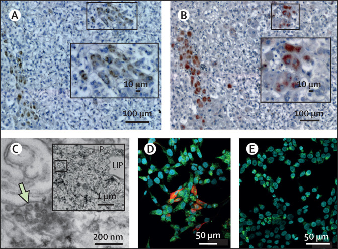
Detection of SARS-CoV-2 in human adrenal gland from a patient who died due to COVID-19 (A) Detection of SARS-CoV-2 RNA by in situ hybridization (ISH; brown DAB staining) and (B) its spike protein by immunohistochemistry (red AEC staining) was found in the same region of two serial 1 μm-thick tissue sections of the adrenal gland. Inserts depict the same regions but were captured at lower magnification showing the chosen area of virus positivity (white squares). Scattered expression of either mRNA or protein can be found in the inner parts of the adrenal gland cortex close to the medulla. Scale bars in pictures from panel A and B represent 100 μm (10 μm in the insert image). (C) Ultrastructural examination of a SARS-CoV-2 triple positive (ISH, immunohistochemistry, and RT-qPCR) adrenal tissue showing numerous viral-like particles in liposome (LIP)-rich adrenocortical cells. A scale bar represents 200 nm. A low magnification picture shown in the insert depicts the region of interest indicated by a green arrow in the enlarged picture. Scale bar in insert indicates 1 μm distance. (D) A positive immunofluorescent red signal (CY3) indicating the expression of spike protein in SARS-CoV-2 infected human adrenocortical cells. (E) Lack of positive signal (CY3) in mock-infected control cells. Adrenocortical cells were additionally stained with an antibody against side-chain cleavage enzyme (CYP11A1), which is a steroidogenic marker (green signal; CY5). Nuclei were counterstained with Hoechst 33342 dye (blue signal). Scale bars in pictures from panel D and E represent 50 μm.
Comment in
-
Letter to the editor of clinical endocrinology: Assessment of adrenal function in patients who survive COVID-19.Clin Endocrinol (Oxf). 2023 Feb;98(2):270-272. doi: 10.1111/cen.14816. Epub 2022 Aug 30. Clin Endocrinol (Oxf). 2023. PMID: 35986449 Free PMC article. No abstract available.
References
-
- Iuga AC, Marboe CC, Yilmaz MM, Lefkowitch JH, Gauran C, Lagana SM. Adrenal vascular changes in COVID-19 autopsies. Arch Pathol Lab Med. 2020;144:1159–1160. - PubMed
Publication types
MeSH terms
LinkOut - more resources
Full Text Sources
Medical
