COVID-19 pneumonia: Relationship between initial chest X-rays and laboratory findings
- PMID: 34801181
- PMCID: PMC8549399
- DOI: 10.1016/j.rxeng.2021.06.003
COVID-19 pneumonia: Relationship between initial chest X-rays and laboratory findings
Abstract
Objective: To analyze the initial findings in chest X-rays of patients with RT-PCR positive for SARS-CoV-2, and to determine whether there is a relationship between the severity of these findings and the clinical and laboratory findings.
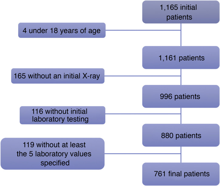
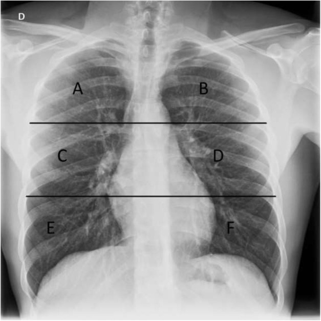
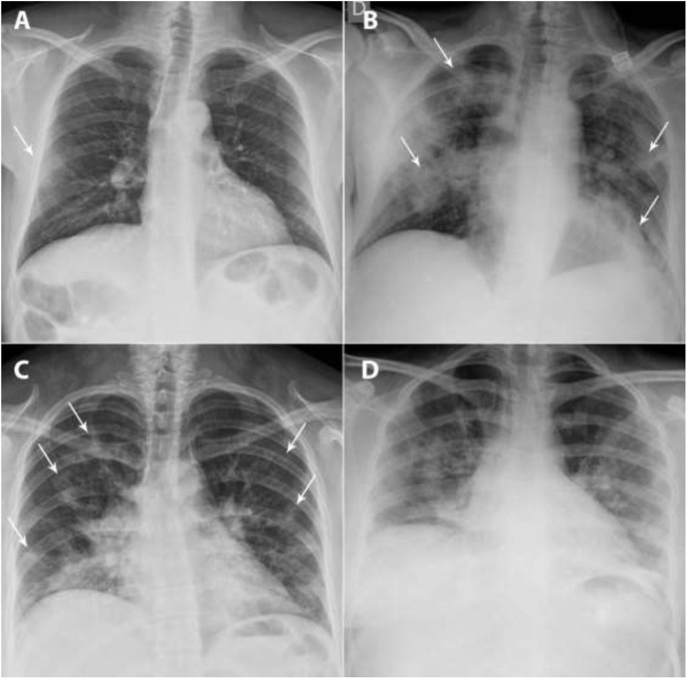
Materials and methods: This retrospective study analyzed the relationship between initial chest X-rays and initial laboratory tests in symptomatic adults with nasopharyngeal RT-PCR results positive for SARS-CoV-2 seen at our center between February 29 and March 23, 2020. Among other radiologic findings, we analyzed ground-glass opacities, consolidations, linear opacities, and pleural effusion. We also used a scale of radiologic severity to assess the distribution and extent of these findings. Among initial laboratory findings, we analyzed leukocytes, lymphocytes, platelets, neutrophil-to-lymphocyte ratio, and C-reactive protein.
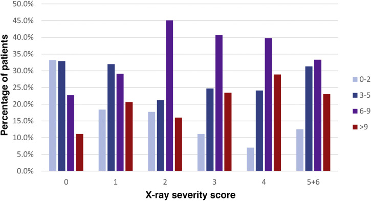
Results: Of 761 symptomatic patients, 639 (84%) required hospitalization and 122 were discharged to their homes. The need for admission increased with increasing scores on the scale of radiologic severity. The extent of initial lung involvement was significantly associated with the laboratory parameters analyzed (P<.05 for platelets, P<.01 for lymphocytes, and P<.001 for the remaining parameters), as well as with the time from the onset of symptoms (P<.001).
Conclusion: It can be useful to use a scale of radiologic severity to classify chest X-ray findings in diagnosing patients with COVID-19, because the greater the radiologic severity, the greater the need for hospitalization and the greater the alteration in laboratory parameters.
Objetivo: Analizar los hallazgos radiológicos iniciales en las radiografías de tórax de pacientes con RT-PCR positiva para SARS-CoV-2 y valorar si existe una relación entre la graduación de los mismos y los datos clínicos y analíticos.
Materiales y métodos: Estudio retrospectivo donde se analizaron las radiografías de tórax iniciales de pacientes adultos sintomáticos entre el 29 de febrero y el 23 de marzo de 2020 con una prueba RT-PCR nasofaríngea positiva para SARS-CoV-2 y una analítica inicial que incluía: leucocitos, linfocitos, plaquetas, cociente linfocitos/leucocitos y PCR. Entre otros hallazgos radiológicos se valoraron: opacidades en vidrio deslustrado, consolidaciones, opacidades lineales y derrame pleural. También la distribución y la extensión de estos hallazgos mediante un índice de gravedad radiográfico.
Resultados: De 761 pacientes sintomáticos, 639 precisaron ingreso hospitalario (84%) y 122 fueron dados de alta con aislamiento domiciliario. La necesidad de ingreso fue mayor cuanto más alto el índice de gravedad radiográfico. Existió una relación estadísticamente significativa entre la extensión de la afectación pulmonar inicial y los parámetros analíticos estudiados (P < ,05 para plaquetas, P < ,01 para linfocitos y P < ,001 para el resto), así como con los días de evolución desde el inicio de los síntomas (P < ,001).
Conclusión: La graduación de los hallazgos radiológicos al diagnóstico y su relación con los datos analíticos podría ser útil a la hora de valorar la evolución de pacientes con COVID-19, pues a mayor índice de gravedad radiográfico, mayor incidencia de ingreso hospitalario y parámetros analíticos más alterados.
Keywords: COVID-19; Chest radiography; Laboratory findings; Puntuación radiográfica; Radiografía de tórax; Radiographic score; Resultados de laboratorio; SARS-CoV-2.
Copyright © 2021 SERAM. Published by Elsevier España, S.L.U. All rights reserved.
Figures
Similar articles
-
COVID-19 pneumonia: relationship between initial chest X-rays and laboratory findings.Radiologia (Engl Ed). 2021 Jun 23;63(6):484-94. doi: 10.1016/j.rx.2021.06.001. Online ahead of print. Radiologia (Engl Ed). 2021. PMID: 34253334 Free PMC article. English, Spanish.
-
Pediatric chest X-rays during the COVID-19 pandemic.Radiologia (Engl Ed). 2021 Mar-Apr;63(2):106-114. doi: 10.1016/j.rx.2020.11.008. Epub 2021 Jan 20. Radiologia (Engl Ed). 2021. PMID: 33483143 Free PMC article.
-
Initial findings in chest X-rays as predictors of worsening lung infection in patients with COVID-19: correlation in 265 patients.Radiologia (Engl Ed). 2021 Jul-Aug;63(4):324-333. doi: 10.1016/j.rxeng.2021.03.006. Epub 2021 Jun 5. Radiologia (Engl Ed). 2021. PMID: 34246423 Free PMC article.
-
Thoracic imaging tests for the diagnosis of COVID-19.Cochrane Database Syst Rev. 2020 Sep 30;9:CD013639. doi: 10.1002/14651858.CD013639.pub2. Cochrane Database Syst Rev. 2020. Update in: Cochrane Database Syst Rev. 2020 Nov 26;11:CD013639. doi: 10.1002/14651858.CD013639.pub3. PMID: 32997361 Updated.
-
CT presentations of adult and pediatric SARS-CoV-2 patients: A review of early COVID-19 data.Radiologia (Engl Ed). 2021 Nov-Dec;63(6):495-504. doi: 10.1016/j.rxeng.2021.04.004. Epub 2021 Sep 4. Radiologia (Engl Ed). 2021. PMID: 34801182 Free PMC article. Review.
Cited by
-
Factors Associated with the Antibiotic Treatment of Children Hospitalized for COVID-19 during the Lockdown in Serbia.Int J Environ Res Public Health. 2022 Nov 24;19(23):15590. doi: 10.3390/ijerph192315590. Int J Environ Res Public Health. 2022. PMID: 36497665 Free PMC article.
-
Chest X-ray Severity Score as a Putative Predictor of Clinical Outcome in Hospitalized Patients: An Experience From a Vietnamese COVID-19 Field Hospital.Cureus. 2022 Mar 19;14(3):e23323. doi: 10.7759/cureus.23323. eCollection 2022 Mar. Cureus. 2022. PMID: 35464539 Free PMC article.
-
Brixia Chest X-ray Score, Laboratory Parameters and Vaccination Status for Prediction of Mortality in COVID-19 Hospitalized Patients.Diagnostics (Basel). 2023 Jun 20;13(12):2122. doi: 10.3390/diagnostics13122122. Diagnostics (Basel). 2023. PMID: 37371019 Free PMC article.
References
-
- Coronavirus disease (COVID-19) – World Health Organization [Internet]. [Accessed 19 Dec 2020]. Available from: https://www.who.int/emergencies/diseases/novel-coronavirus-2019.
-
- WHO Western Pacific | World Health Organization [Internet]. [19 Dec 2020]. Available from: https://www.who.int/westernpacific/emergencies/covid-19.
-
- Actualizacion_274_COVID-19.pdf [Internet]. [19 Dec 2020]. Available from: https://www.mscbs.gob.es/profesionales/saludPublica/ccayes/alertasActual....
MeSH terms
LinkOut - more resources
Full Text Sources
Medical
Research Materials
Miscellaneous