

Multilayer Macula Vessel Density and Visual Field Progression in Glaucoma
- PMID: 34801510
- PMCID: PMC9469932
- DOI: 10.1016/j.ajo.2021.11.018
Multilayer Macula Vessel Density and Visual Field Progression in Glaucoma
Abstract
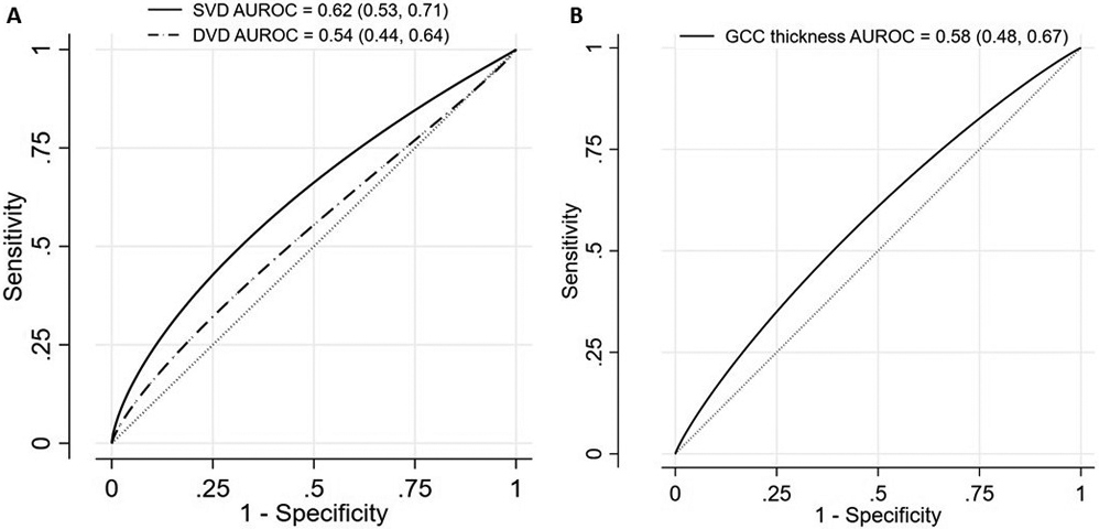
Purpose: To evaluate the association of macular superficial vessel density (SVD) and projection-resolved deep vessel density (DVD) with past visual field (VF) progression in patients with primary open-angle glaucoma.
Design: Retrospective cohort.
Methods: In this longitudinal study, 208 eyes of 147 patients with glaucoma from the Diagnostics Innovations in Glaucoma Study were included. Eligible participants were required to have at least five 24-2 VF tests over a minimum follow-up period of 3 years before macular optical coherence tomography angiography imaging. VF progression was defined based on both event-based pointwise linear regression and trend-based methods. The association of macular SVD and DVD with the probability and rate of past VF progression was evaluated using a linear mixed effects model.
Results: Fifty-two (25%) eyes had VF progression based on the pointwise linear regression based criterion at the end of a mean ± standard deviation follow-up duration of 6.9 ± 1.2 years. In the event-based multivariable analysis, a lower baseline SVD was associated with a higher likelihood of past VF progression (odds ratio per 1% lower. 1.28; 95% confidence interval, 1.02-1.59). Similarly, in the trend-based multivariable analysis, lower macular SVD was associated with a faster past rate of mean deviation decline (coefficient = -0.03 dB/year; 95% confidence interval, -0.04 to -0.01). Event-based and trend-based analyses found no significant associations for macular DVD with the likelihood/rate of past VF progression (P > .05).
Conclusions: Lower macular SVD, and not DVD, was associated with a higher probability of past VF progression. Macular optical coherence tomography angiography imaging shows promise for identifying eyes at risk of VF progression in patients with glaucoma.
Copyright © 2021 Elsevier Inc. All rights reserved.
Figures
References
-
- Weinreb RN, Leung CK, Crowston JG, et al. Primary open-angle glaucoma. 2016;2:1–19. - PubMed
-
- Kwon JM, Weinreb RN, Zangwill LM, Suh MH. Parapapillary Deep-Layer Microvasculature Dropout and Visual Field Progression in Glaucoma. Am J Ophthalmol 2019;200:65–75. - PubMed
-
- Gordon MO, Beiser JA, Brandt JD, et al. The Ocular Hypertension Treatment Study: baseline factors that predict the onset of primary open-angle glaucoma. 2002;120:714–720. - PubMed
Publication types
MeSH terms
Grants and funding
LinkOut - more resources
Full Text Sources
Medical
Miscellaneous
