. 2021 Dec;297(6):101428.
doi: 10.1016/j.jbc.2021.101428. Epub 2021 Nov 19.

Development of a versatile HPLC-based method to evaluate the activation status of small GTPases

Affiliations

Development of a versatile HPLC-based method to evaluate the activation status of small GTPases

Makoto Araki et al. J Biol Chem. 2021 Dec.

Abstract

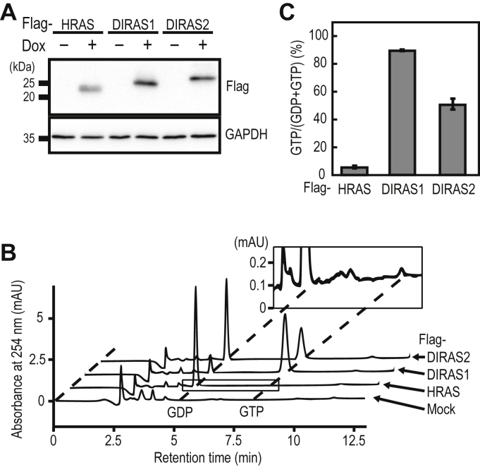
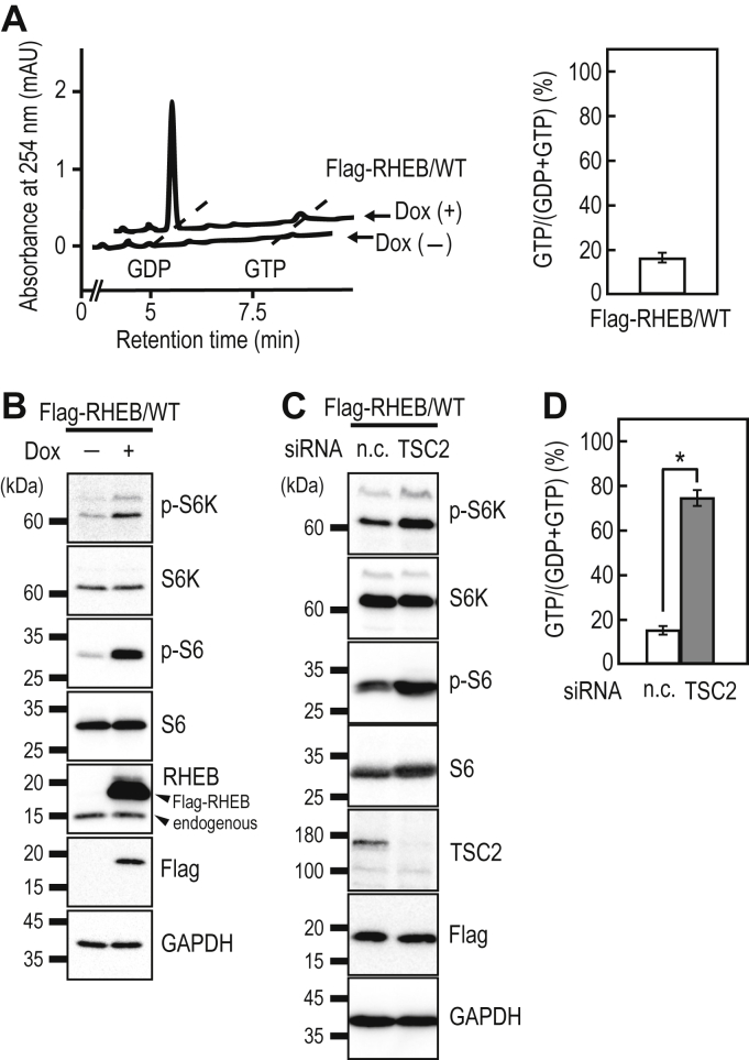
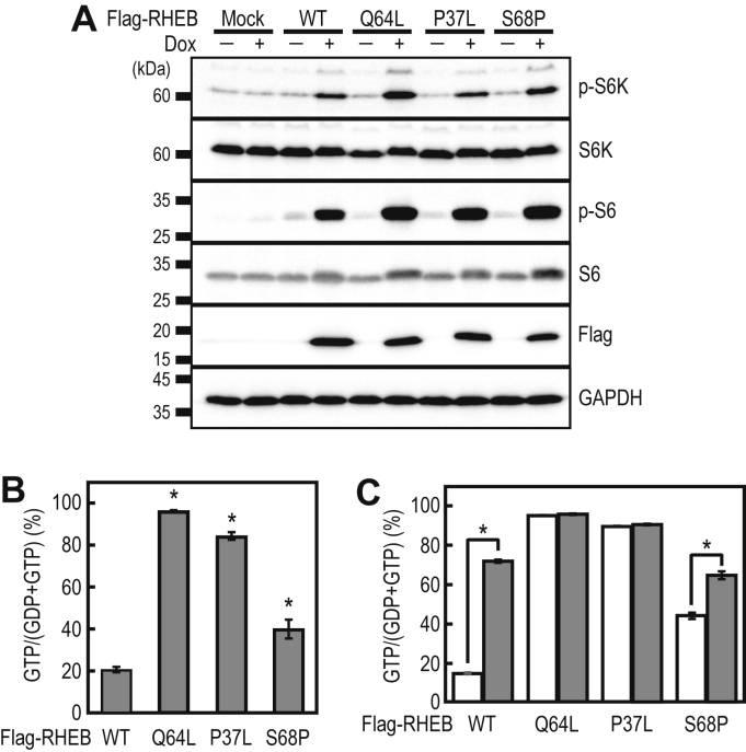
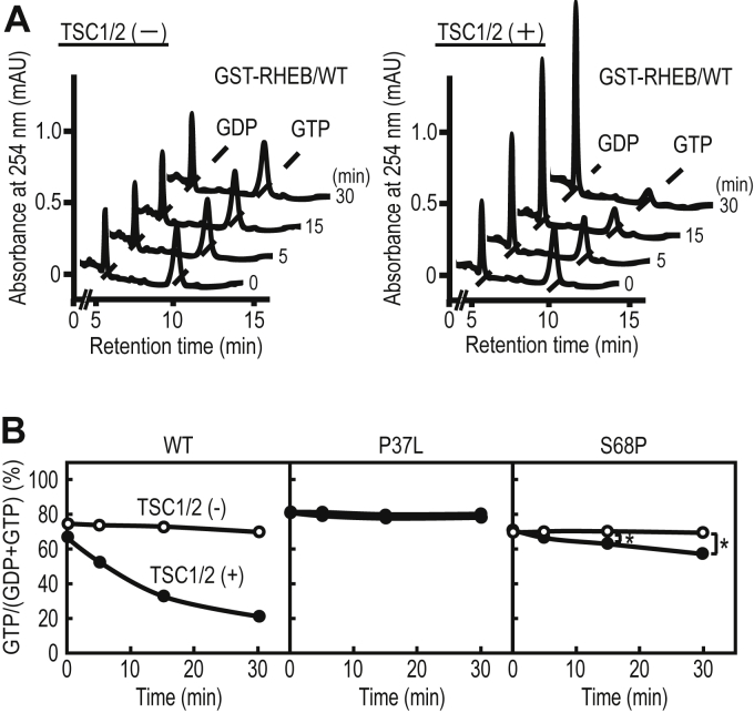
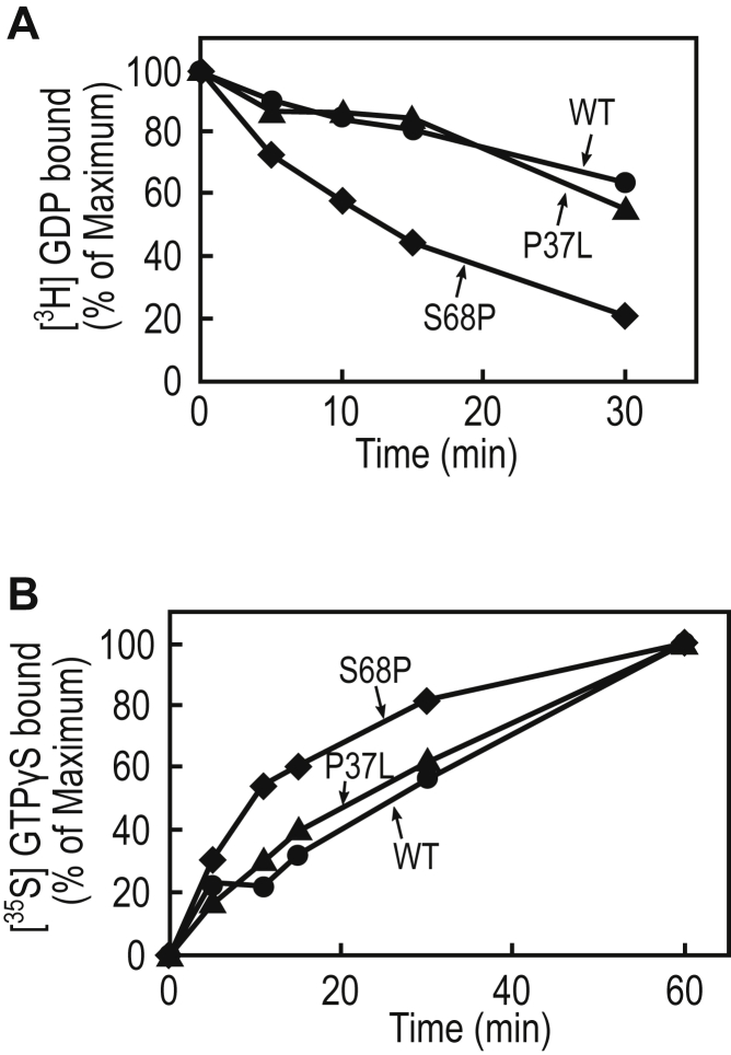
Small GTPases cycle between an inactive GDP-bound and an active GTP-bound state to control various cellular events, such as cell proliferation, cytoskeleton organization, and membrane trafficking. Clarifying the guanine nucleotide-bound states of small GTPases is vital for understanding the regulation of small GTPase functions and the subsequent cellular responses. Although several methods have been developed to analyze small GTPase activities, our knowledge of the activities for many small GTPases is limited, partly because of the lack of versatile methods to estimate small GTPase activity without unique probes and specialized equipment. In the present study, we developed a versatile and straightforward HPLC-based assay to analyze the activation status of small GTPases by directly quantifying the amounts of guanine nucleotides bound to them. This assay was validated by analyzing the RAS-subfamily GTPases, including HRAS, which showed that the ratios of GTP-bound forms were comparable with those obtained in previous studies. Furthermore, we applied this assay to the investigation of psychiatric disorder-associated mutations of RHEB (RHEB/P37L and RHEB/S68P), revealing that both mutations cause an increase in the ratio of the GTP-bound form in cells. Mechanistically, loss of sensitivity to TSC2 (a GTPase-activating protein for RHEB) for RHEB/P37L, as well as both decreased sensitivity to TSC2 and accelerated guanine-nucleotide exchange for RHEB/S68P, is involved in the increase of their GTP-bound forms, respectively. In summary, the HPLC-based assay developed in this study provides a valuable tool for analyzing small GTPases for which the activities and regulatory mechanisms are less well understood.

Keywords: GTPase-activating protein; RHEB; Ras protein; high-performance liquid chromatography; mammalian target of rapamycin; signal transduction; small GTPase; tuberous sclerosis complex.


Conflict of interest statement

Conflict of interest The authors declare that they have no conflicts of interest with the contents of this article.

Figures

Figure 1
Figure 1
Separation of adenine and guanine nucleotides by IP-RP-HPLC. Representative chromatogram of a standard mixture of four nucleotides (ADP, ATP, GDP, and GTP, each 10 pmol) using IP-RP-HPLC. The nucleotides were detected by a UV detector at a wavelength of 254 nm. The peaks of GDP, ADP, GTP, and ATP were detected at 5.4, 6.2, 8.6, and 10.9 min, respectively, on chromatograms. IP-RP-HPLC, ion-pair reversed-phase HPLC.
Figure 2
Figure 2
Analysis of the guanine-nucleotide-bound forms of RAS-family GTPases in HeLa cells.A, Dox-dependent expression of Flag-tagged HRAS, DIRAS1, and DIRAS2 in HeLa cells. The cell lines expressing the indicated proteins in a Dox-dependent manner were cultured in the absence or presence of 1 μg/ml of Dox for 24 h, and the cell lysates were subjected to Western blot analysis using anti-Flag and anti-GAPDH antibodies. B, representative chromatogram of guanine nucleotides bound to Flag-tagged HRAS, DIRAS1, and DIRAS2 in HeLa cells. Anti-Flag immunoprecipitates from the indicated cell lines were subjected to IP-RP-HPLC analysis. To visualize the GTP signal of HRAS, a magnified view of the enclosed area of HRAS is shown as the inset. C, the relative amounts of guanine nucleotides associated with Flag-tagged proteins were quantified from the peak areas of GDP and GTP using IP-RP-HPLC. The data are the means ± SD from three independent experiments. IP-RP-HPLC, ion-pair reversed-phase HPLC; Dox, doxycycline.
Figure 3
Figure 3
Analysis of the activation status of Flag-RHEB in HeLa cells.A, cell lines expressing Flag-RHEB/WT in a Dox-dependent manner were cultured without or with 1 μg/ml of Dox for 24 h, and anti-Flag immunoprecipitates from the cell lysates were subjected to IP-RP-HPLC analysis. Representative chromatogram of guanine nucleotides bound to Flag-RHEB/WT in HeLa cells (left panel). The relative amounts of guanine nucleotides associated with Flag-RHEB/WT were quantified from the peak areas of GDP and GTP (right panel). The data are the means ± SD from three independent experiments. B, cell lines expressing Flag-RHEB/WT in a Dox-dependent manner were cultured without or with 1 μg/ml of Dox for 24 h, and the cell lysates were subjected to Western blot analysis using the indicated antibodies. C and D, effect of TSC2 knockdown on mTORC1 signaling and the guanine-nucleotide bound state of Flag-RHEB/WT. The cell lines expressing Flag-RHEB/WT in a Dox-dependent manner were transfected with negative control (n.c.) or TSC2 siRNAs and cultured for 48 h, followed by 24 h of culture in the presence of 1 μg/ml of Dox. The cell lysates were subjected to Western blot analysis (C) or IP-RP-HPLC analysis (D). The data are the means ± SD from three independent experiments. ∗ p < 0.05, Student’s t test. Dox, doxycycline; IP-RP-HPLC, ion-pair reversed-phase HPLC; mTORC1, mechanistic target of rapamycin complex 1; RHEB, Ras homolog enriched in brain.
Figure 4
Figure 4
Analysis of the activation status of the disease-associated RHEB mutants in HeLa cells.A, the cell lines expressing Flag-RHEB/WT or mutants in a Dox-dependent manner were cultured for 24 h in the absence or presence of 1 μg/ml of Dox, and the cell lysates were subjected to Western blot analysis using the indicated antibodies. B, anti-Flag immunoprecipitates from the cell lines expressing Flag-RHEB/WT and mutants were subjected to IP-RP-HPLC analysis. The data are the means ± SD from three independent experiments. ∗ p < 0.05, compared with WT by Dunnett’s multiple comparison test. C, cell lines expressing Flag-RHEB/WT and mutants in a Dox-dependent manner were transfected with negative control (open bars) or TSC2 (closed bars) siRNAs and cultured for 48 h, after 24 h of culture in the presence of 1 μg/ml of Dox. The cell lysates were subjected to anti-Flag immunoprecipitation for IP-RP-HPLC analysis. The data are the means ± SD from three independent experiments. ∗ p < 0.05, Student’s t test. IP-RP-HPLC, ion-pair reversed-phase HPLC; RHEB, Ras homolog enriched in brain.
Figure 5
Figure 5
Effect of the TSC1/2 complex on the GTPase activity of WT and mutant RHEB proteins.A, representative chromatograms of the GTPase assay using IP-RP-HPLC. The GST-fusion proteins of WT RHEB were incubated at 30 °C without (left panel) or with (right panel) Flag-TSC1/2 immunoprecipitation beads at the indicated times, and the aliquots were subjected to IP-RP-HPLC analysis. B, the GST-fusion proteins of the WT and mutant forms of RHEB were incubated at 30 °C without (open circle) or with (closed circle) Flag-TSC1/2 immunoprecipitation beads at the indicated times, and the aliquots were subjected to IP-RP-HPLC analysis. The data are the means from three independent experiments. The error bars are not shown as the standard deviations of the data were within 1%. ∗ p < 0.05, Student’s t test. IP-RP-HPLC, ion-pair reversed-phase HPLC; RHEB, Ras homolog enriched in brain.
Figure 6
Figure 6
Analysis of the GDP-dissociation and GTPγS-association rates of WT and mutant RHEB proteins.A, GDP-dissociation assay. The GST-fusion proteins of RHEB/WT (circles), P37L (triangles), or S68P (rhombuses) were incubated with 5 μM [3H]GDP in the presence of 5 mM Mg2+ for 20 min, and unlabeled GTPγS (200 μM) was added to the reaction mixture. At the indicated times, the aliquots (20 μl) were withdrawn and analyzed for [3H]GDP binding, as described in the “Experimental procedures.” The data are the means from two independent experiments. B, GTPγS-binding assay. The GST-fusion proteins of RHEB/WT (circles), P37L (triangles), or S68P (rhombuses) were incubated with 5 μM [35S]GTPγS for the indicated times, and [35S]GTPγS binding was determined, as described in the “Experimental procedures.” The data are the means from two independent experiments. RHEB, Ras homolog enriched in brain.

References

    1. Colicelli J. Human RAS superfamily proteins and related GTPases. Sci. STKE. 2004;2004:RE13. - PMC - PubMed
    1. Wennerberg K., Rossman K.L., Der C.J. The Ras superfamily at a glance. J. Cell Sci. 2005;118:843–846. - PubMed
    1. Cherfils J., Zeghouf M. Regulation of small GTPases by GEFs, GAPs, and GDIs. Physiol. Rev. 2013;93:269–309. - PubMed
    1. Boulter E., Garcia-Mata R., Guilluy C., Dubash A., Rossi G., Brennwald P.J., Burridge K. Regulation of Rho GTPase crosstalk, degradation and activity by RhoGDI1. Nat. Cell Biol. 2010;12:477–483. - PMC - PubMed
    1. Müller M.P., Goody R.S. Molecular control of Rab activity by GEFs, GAPs and GDI. Small GTPases. 2017;9:5–21. - PMC - PubMed

Publication types

MeSH terms

Substances