. 2021 Dec;297(6):101423.
doi: 10.1016/j.jbc.2021.101423. Epub 2021 Nov 18.

Rescuing activity of oxygen-damaged pyruvate formate-lyase by a spare part protein

Affiliations

Rescuing activity of oxygen-damaged pyruvate formate-lyase by a spare part protein

Mary C Andorfer et al. J Biol Chem. 2021 Dec.

Abstract

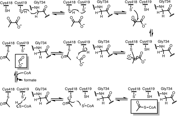
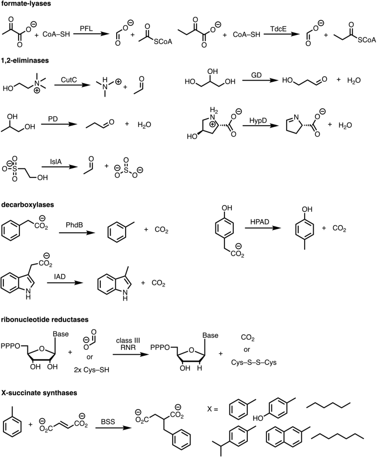
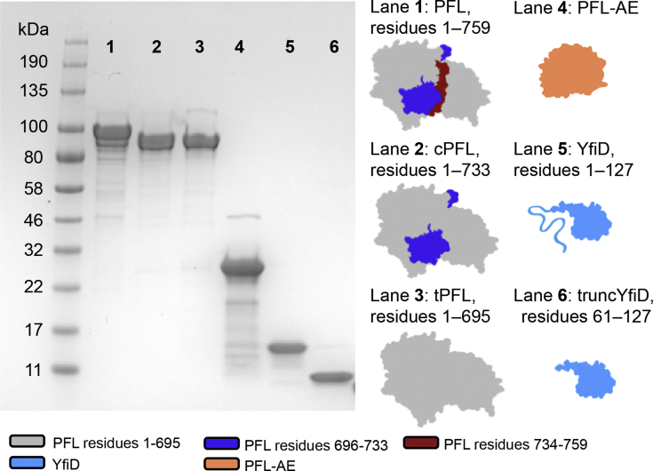
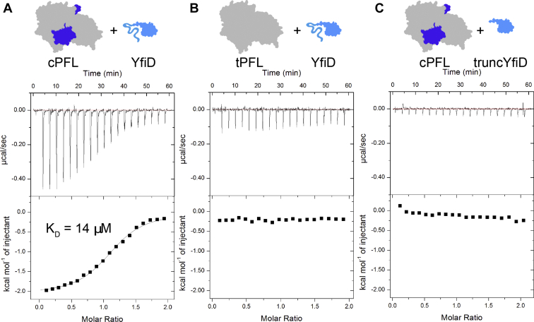
Pyruvate formate-lyase (PFL) is a glycyl radical enzyme (GRE) that converts pyruvate and coenzyme A into acetyl-CoA and formate in a reaction that is crucial to the primary metabolism of many anaerobic bacteria. The glycyl radical cofactor, which is posttranslationally installed by a radical S-adenosyl-L-methionine (SAM) activase, is a simple and effective catalyst, but is also susceptible to oxidative damage in microaerobic environments. Such damage occurs at the glycyl radical cofactor, resulting in cleaved PFL (cPFL). Bacteria have evolved a spare part protein termed YfiD that can be used to repair cPFL. Previously, we obtained a structure of YfiD by NMR spectroscopy and found that the N-terminus of YfiD was disordered and that the C-terminus of YfiD duplicates the structure of the C-terminus of PFL, including a β-strand that is not removed by the oxygen-induced cleavage. We also showed that cPFL is highly susceptible to proteolysis, suggesting that YfiD rescue of cPFL competes with protein degradation. Here, we probe the mechanism by which YfiD can bind and restore activity to cPFL through enzymatic and spectroscopic studies. Our data show that the disordered N-terminal region of YfiD is important for YfiD glycyl radical installation but not for catalysis, and that the duplicate β-strand does not need to be cleaved from cPFL for YfiD to bind. In fact, truncation of this PFL region prevents YfiD rescue. Collectively our data suggest the molecular mechanisms by which YfiD activation is precluded both when PFL is not damaged and when it is highly damaged.

Keywords: bacterial metabolism; cofactor repair; electron paramagnetic resonance (EPR) spectroscopy; enzyme inactivation; glycyl radical enzyme; isothermal titration calorimetry (ITC); oxygen-sensitive enzymes; protein complex; radical chemistry; spare part protein.


Conflict of interest statement

Conflicts of interest The authors declare that they have no conflicts of interest with the contents of this article.

Figures

Figure 1
Figure 1
Proposed mechanism of PFL.
Figure 2
Figure 2
GREs catalyze a wide variety of challenging reactions using radical chemistry.
Figure 3
Figure 3
Initial model for O2-damaged PFL rescue of activity by YfiD and topology diagrams.A, crystal structures of PFL and PFL-AE (PDB ID: 2PFL and 3CB8, respectively) and NMR structure of YfiD (PDB ID: 6OWR) were used to create cartoons. No structural data are available for any of the above protein complexes—cartoons of complexes were created by manually docking structures as previously described. Color coding is as follows: PFL residues 1 to 695 in gray, PFL residues 696 to 733 in dark blue, PFL residues 734 to 759 in red, PFL-AE in orange, YfiD in light blue. B, topology diagram of PFL with residues 1 to 695 in gray, 696 to 733 in dark blue, and 734 to 759 in red. C, topology diagram of truncated PFL (tPFL, gray) in complex with YfiD (light blue). Residues 1 to 60 of YfiD are disordered in the NMR structure and residues 61 to 127 of YfiD have the same fold as residues 693 to 759 of PFL, which includes a β strand (β10 in PFL and y-β1 in YfiD), the glycyl radical loop, and a C-terminal helix.
Figure 4
Figure 4
Summary of constructs. Gel, cartoons, names, and construct length for all proteins used in this study.
Figure 5
Figure 5
Glycyl radical installation comparisons.Top: Reaction conditions for activations of PFL and PFL:YfiD complexes. Middle: Cartoon representations of PFL and PFL:YfiD complexes. Bottom: EPR data used to quantify amounts of glycyl radical (N = 3). Briefly, in an anaerobic chamber, PFL variants (200 μM final conc.) and YfiD variants (200 μM final conc.) were diluted with 20 mM HEPES pH 7.2 to a final volume of 150 μl. Pyruvate, PFL-AE, AdoMet, and 5-deazariboflavin were added to each reaction. Activation buffer was added to each reaction for a final volume of 300 μl. The activations were mixed by pipetting and illuminated using a 500 W halogen lamp for 15 to 30 min. EPR spectroscopy was used to quantify glycyl radical content. EPR parameters were as follows: 80 K, 9.37 GHz, modulation amplitude of 3 G, microwave power of 1.26 μW.
Figure 6
Figure 6
Saturation plots for wild-type PFL and four PFL:YfiD complexes. Production of acetyl-CoA by PFL and PFL:YfiD complexes was measured through a coupled assay with citrate synthase and malic acid dehydrogenase. Inside of an anaerobic chamber, citrate synthase (6 U per reaction), malic acid dehydrogenase (14 U per reaction), CoA (2.5–400 μM) were added to assay buffer (150 mM Tris pH 8.5, 10 mM L-malate, 10 mM pyruvate, 3 mM NAD). Activated PFL or PFL:YfiD mixture was added to initiate the reaction and immediately pipetted to mix. Data were collected on an Ocean Optics Spectrometer at 366 nm to measure absorbance of NADH. Initial velocity curves were conducted in triplicate for each CoA concentration at 21 °C and plotted using Prism nonlinear regression software to calculate KM and Vmax for each complex. EPR spectroscopy was used to measure glycyl radical content for PFL and PFL:YfiD complexes, and the final concentrations of radical in reactions were used as Etot. Vmax and Etot were used to calculate kcat.
Figure 7
Figure 7
ITC binding data for YfiD and truncYfiD added as titrant to cPFL and tPFL.A, an exothermic binding event between cPFL and YfiD occurs. The best fit is consistent with a KD of 14 μM for the cPFL:YfiD complex. Initial [cPFL] in cell = 224 μM (186 μM final conc.), initial [YfiD] = 2.129 mM (361 μM final conc.). B, no clear binding event between tPFL and YfiD is observed. Instead, only the heat of dilution for YfiD can be observed. Initial [tPFL] in cell = 224 μM (186 μM final conc.), initial [YfiD] = 2.1 mM (356 μM final conc.). C, no clear, enthalpic binding event between cPFL and truncYfiD is observed. Instead, only the heat of dilution for truncYfiD can be observed. Initial [cPFL] in cell = 173 μM (143 μM final conc.), initial [truncYfiD] = 1.73 mM (294 μM final conc.).
Figure 8
Figure 8
Revised model for YfiD rescue of O2-damaged PFL. Crystal structures of PFL and PFL-AE (PDB ID: 2PFL and 3CB8, respectively) and NMR structure of YfiD (PDB ID: 6OWR) were used to create cartoons. No structural data are available for any of the above protein complexes—cartoons of complexes were created by manually docking structures as previously described. Color coding is as follows: PFL residues 1 to 695 in gray, PFL residues 696 to 733 in dark blue, PFL residues 734 to 759 in red, PFL-AE in orange, YfiD in light blue.

References

    1. Backman L.R.F., Funk M.A., Dawson C.D., Drennan C.L. New tricks for the glycyl radical enzyme family. Crit. Rev. Biochem. Mol. Biol. 2017;52:674–695. - PMC - PubMed
    1. Sun X., Ollagnier S., Schmidt P.P., Atta M., Mulliez E., Lepape L., Eliasson R., Graslund A., Fontecave M., Reichard P., Sjoberg B.M. The free radical of the anaerobic ribonucleotide reductase from Escherichia coli is at glycine 681. J. Biol. Chem. 1996;271:6827–6831. - PubMed
    1. Leuthner B., Leutwein C., Schulz H., Horth P., Haehnel W., Schiltz E., Schagger H., Heider J. Biochemical and genetic characterization of benzylsuccinate synthase from Thauera aromatica: A new glycyl radical enzyme catalysing the first step in anaerobic toluene metabolism. Mol. Microbiol. 1998;28:615–628. - PubMed
    1. Levin B.J., Balskus E.P. Discovering radical-dependent enzymes in the human gut microbiota. Curr. Opin. Chem. Biol. 2018;47:86–93. - PubMed
    1. Levin B.J., Huang Y.Y., Peck S.C., Wei Y., Martinez-Del Campo A., Marks J.A., Franzosa E.A., Huttenhower C., Balskus E.P. A prominent glycyl radical enzyme in human gut microbiomes metabolizes trans-4-hydroxy-l-proline. Science. 2017;355 - PMC - PubMed

Publication types

MeSH terms

LinkOut - more resources