Review
. 2022 Jan:180:114043.
doi: 10.1016/j.addr.2021.114043. Epub 2021 Nov 18.

Towards controlled drug delivery in brain tumors with microbubble-enhanced focused ultrasound

Affiliations
Review

Towards controlled drug delivery in brain tumors with microbubble-enhanced focused ultrasound

Scott Schoen Jr et al. Adv Drug Deliv Rev. 2022 Jan.

Abstract

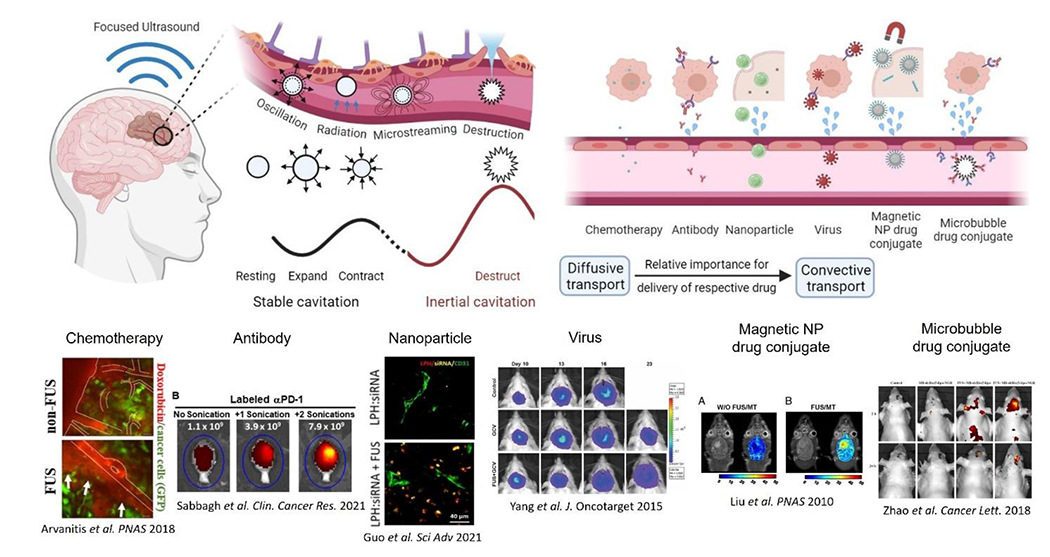
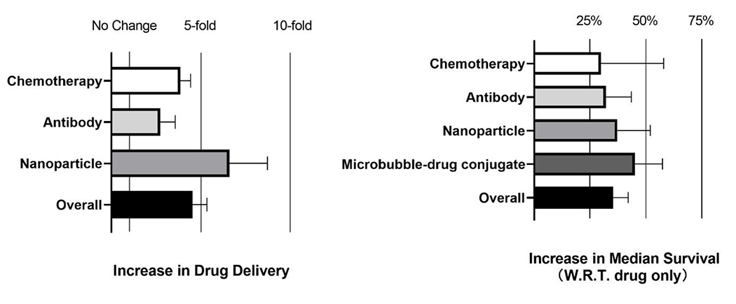
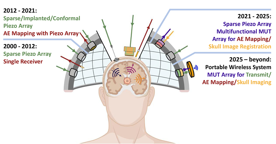
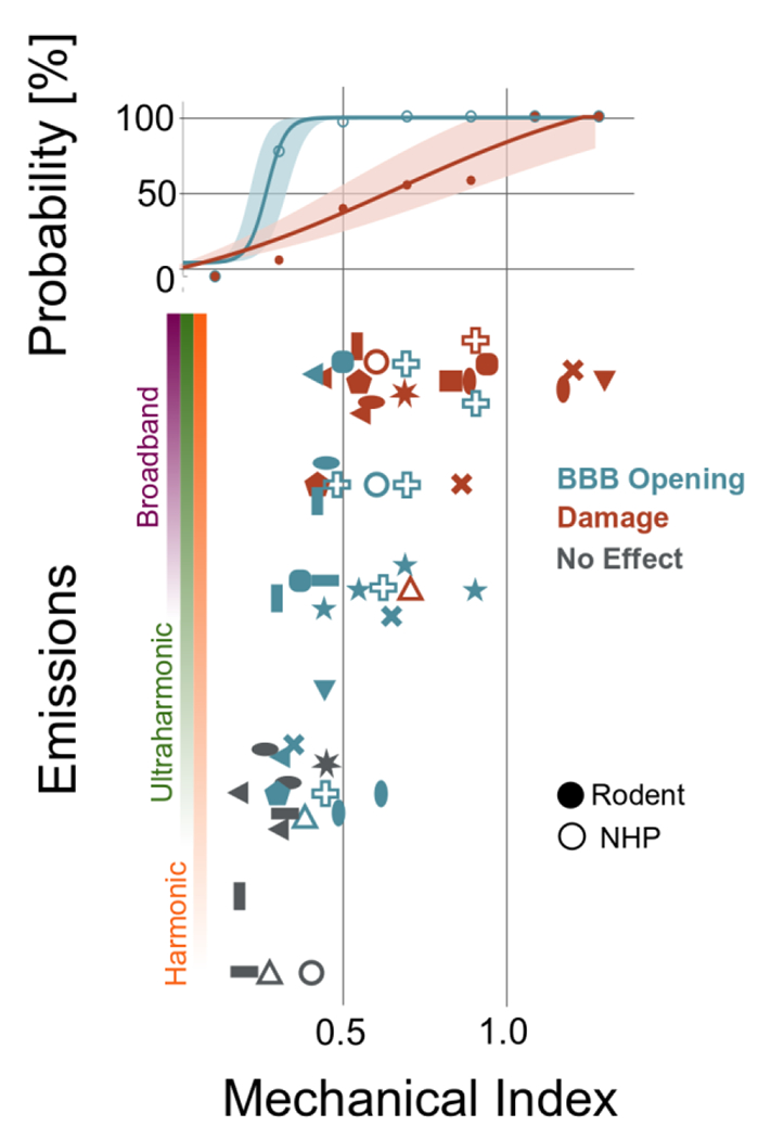
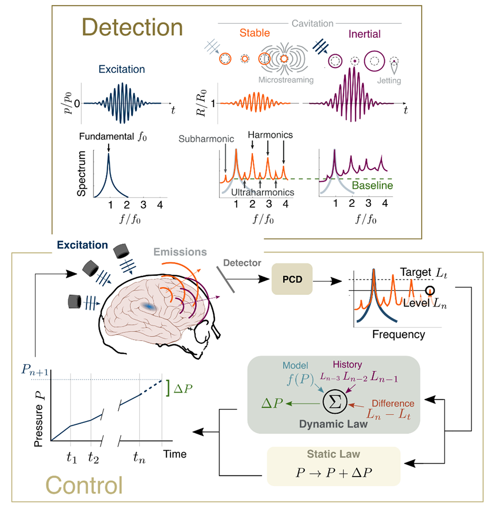
Brain tumors are particularly challenging malignancies, due to their location in a structurally and functionally distinct part of the human body - the central nervous system (CNS). The CNS is separated and protected by a unique system of brain and blood vessel cells which together prevent most bloodborne therapeutics from entering the brain tumor microenvironment (TME). Recently, great strides have been made through microbubble (MB) ultrasound contrast agents in conjunction with ultrasound energy to locally increase the permeability of brain vessels and modulate the brain TME. As we elaborate in this review, this physical method can effectively deliver a wide range of anticancer agents, including chemotherapeutics, antibodies, and nanoparticle drug conjugates across a range of preclinical brain tumors, including high grade glioma (glioblastoma), diffuse intrinsic pontine gliomas, and brain metastasis. Moreover, recent evidence suggests that this technology can promote the effective delivery of novel immunotherapeutic agents, including immune check-point inhibitors and chimeric antigen receptor T cells, among others. With early clinical studies demonstrating safety, and several Phase I/II trials testing the preclinical findings underway, this technology is making firm steps towards shaping the future treatments of primary and metastatic brain cancer. By elaborating on its key components, including ultrasound systems and MB technology, along with methods for closed-loop spatial and temporal control of MB activity, we highlight how this technology can be tuned to enable new, personalized treatment strategies for primary brain malignancies and brain metastases.

Keywords: Blood brain barrier; Blood tumor barrier; Brain cancer; Drug delivery; Focused ultrasound; Micro-bubbles; Ultrasound Immunomodulation.


Conflict of interest statement

Declaration of Competing Interest The authors declare that they have no known competing financial interests or personal relationships that could have appeared to influence the work reported in this paper.

Figures

Figure 1:
Figure 1:
Circulating microbubble contrast agents are excited by transcranial focused ultrasound. The resulting oscillations give rise to various mechanical effects including microstreaming, radiation forces, and destruction via shockwaves and jetting during bubble collapse. Such effects alter the permeability of the blood brain barrier and enable more convective transport of larger therapeutic agents.
Figure 2.
Figure 2.
Analysis of increase in drug delivery (left) and percentage increase in median survival time (IST) (right) after MB-FUS delivery of anticancer agents in murine brain tumor models. Reported increase is with respect to drug only group. All data and citations used to create the plots are provided in Suppl. Table 1.
Figure 3.
Figure 3.
Evolution of FUS and emissions monitoring transducer technology and potential future implementations. Color Coding: Green: Incident ultrasound, Red: Recorded acoustic emissions (AE), Gold: Skull imaging with ultrasound, Purple: Recorded AE and skull imaging with the same transducer.
Figure 4
Figure 4
Emissions and bioeffects reported during MB-FUS for BBB opening. Vertical axis quantifies the type of emissions observed at the given mechanical index (horizontal axis). Blue markers indicate BBB-opening while orange indicate observed tissue damage; the colored bars indicate the probability of no effect, BBB opening, and damage among the studies at that particular MI. Solid markers represent measurements for rodents (mouse, rat, or rabbit) while hollow markers indicate non-human primate studies. Full data provided in Suppl. Table 2.
Figure 5.
Figure 5.
Detection and control of cavitation. (Top) The therapeutic ultrasound pulse has fundamental frequency f0. The circulating microbubbles are excited and undergo stable oscillations (inducing microstreaming and radiating harmonic, ultraharmonic, and subharmonic emissions) or, at higher applied pressure magnitudes, transient inertial cavitation (resulting in jetting and broadband emissions). (Bottom) These emissions can be monitored by a passive detector, which adjusts the applied pressure P based on the type and level L of these emissions relative to a target.

Similar articles

Cited by

References

    1. Hawkins BT, Davis TP, The Blood-Brain Barrier/Neurovascular Unit in Health and Disease, Pharmacol. Rev 57 (2005) 173–185. 10.1124/pr.57.2.4. - DOI - PubMed
    1. McConnell HL, Kersch CN, Woltjer RL, Neuwelt EA, The Translational Significance of the Neurovascular Unit*, J. Biol. Chem 292 (2017) 762–770. 10.1074/jbc.R116.760215. - DOI - PMC - PubMed
    1. Phenix CP, Togtema M, Pichardo S, Zehbe I, Curiel L, High Intensity Focused Ultrasound Technology, its Scope and Applications in Therapy and Drug Delivery, J. Pharm. Pharm. Sci 17 (2014) 136–153. 10.18433/J3ZP5F. - DOI - PubMed
    1. Arvanitis CD, Ferraro GB, Jain RK, The blood-brain barrier and blood-tumour barrier in brain tumours and metastases, Nat. Rev. Cancer 20 (2020) 26–41. 10.1038/s41568-019-0205-x. - DOI - PMC - PubMed
    1. Arvanitis CD, Ferraro GB, Jain RK, The blood-brain barrier and blood-tumour barrier in brain tumours and metastases, Nat. Rev. Cancer 20 (2020) 26–41. 10.1038/s41568-019-0205-x. - DOI - PMC - PubMed

Publication types

Substances