Case Reports
. 2022 Jul;26(3):153-159.
doi: 10.7874/jao.2021.00297. Epub 2021 Nov 23.

Spontaneous Upbeat Nystagmus and Selective Anterior Semicircular Canal Hypofunction on Video Head Impulse Test: A New Variant of Canalith Jam?

Affiliations
Case Reports

Spontaneous Upbeat Nystagmus and Selective Anterior Semicircular Canal Hypofunction on Video Head Impulse Test: A New Variant of Canalith Jam?

Andrea Castellucci et al. J Audiol Otol. 2022 Jul.

Abstract

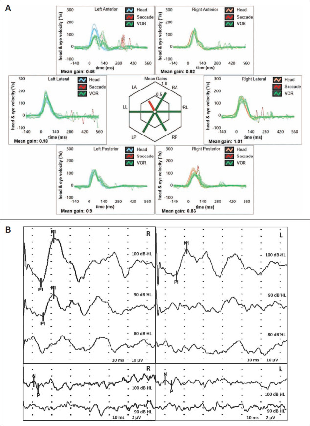
We describe a rare case of spontaneous upbeat nystagmus (UBN) attributable to a canalith jam involving the anterior semicircular canal (ASC) in a patient in whom comprehensive vestibular assessment was useful to identify the underlying pathomechanism. A 56-year-old woman with unsteadiness following repositioning procedures for left-sided benign paroxysmal positional vertigo (BPPV) presented with spontaneous UBN that showed slight right torsional components. A vestibular test battery detected isolated left ASC hypofunction on a video-head impulse test (Video-HIT). We postulated a persistent utriculopetal deflection of the left ASC cupula, which was attributable to entrapment of debris in a narrow canal tract, with consequent sustained inhibition of the ampullary afferents. Although spontaneous UBN receded after impulsive physical therapy, unsteadiness deteriorated into positional vertigo secondary to canalolithiasis involving the ipsilateral posterior canal. In our view, physical therapy possibly fragmented the canalith jam and released free-floating otoconia that eventually settled into the ipsilateral posterior canal. Video HIT revealed normalization of ASC hypofunction, and leftsided posterior canal canalolithiasis was successfully treated using appropriate repositioning procedures. We propose that a canalith jam involving the ASC should be considered in the differential diagnosis of spontaneous UBN, particularly in patients with a history of BPPV and isolated ASC hypofunction detected on video HIT.

Keywords: Benign paroxysmal positional vertigo; Head impulse test; Otoconia; Semicircular canals; Vertical nystagmus.


Figures

Fig. 1.
Fig. 1.
Patient's vestibular assessment at presentation. A: Video-HIT performed using the ICS Impulse device (Otometrics, Natus Medical Inc, Taastrup, Denmark) assessing the VOR-gain (eye velocity/head velocity) for all six semicircular canals. Mean value of VOR-gain is reported for each canal. Gain values are considered normal if >0.8 for lateral canals and >0.7 for vertical canals. A selective reduction for the VOR-gain of the left ASC (0.46) with both overt and covert saccades could be observed. B: Cervical (above) and ocularVEMPs (below) for air-conducted sounds recorded using a 2-channel evoked potential acquisition system (Viking, Nicolet EDX, CareFusion, Heidelberg, Germany). Potentials were recorded delivering tone bursts (frequency: 500 Hz, duration: 8 ms, stimulation rate 5 Hz) via headphones. VEMPs testing revealed slightly asymmetrical amplitudes for both cervical (R: 69.2 µV and L: 41.4 µV at 100 dB HL stimuli) and ocular-VEMPs (R: 2.2 µV and L: 1.5 µV for 100 dB HL sounds) with an asymmetry ratio within normality ranges for both measurements (25% and 19%, respectively). ASC, anterior semicircular canal; HIT, head impulse test; L, left; LA, left anterior; LL, left lateral; LP, left posterior; R, right; RA, right anterior; RL, right lateral; RP, right posterior; VEMPs, vestibular-evoked myogenic potentials; VOR, vestibulo-ocular reflex.
Fig. 2.
Fig. 2.
Imaging and supposed pathomechanism for presenting symptoms and signs. A: Coronal images of temporal bone HRCT scan, with parasagittal reconstructions along the Pöschl planes, showing normal bony roof overlying the left ASC (white arrowhead). B: Schematic drawing of the left membranous labyrinth showing the assumed mechanism for presenting spontaneous UBN with subtle right torsional components and for selective left ASC hypofunction. A debris clot entrapped within the left ASC is supposed to generate a continuous positive pressure (black dashed arrow) on the cupula, inhibiting ampullary afferents and preventing dynamic responses for the canal by blocking endolymphatic flows. ASC-cupula is bent utriculopetally (red line) with respect to its original position (black dashed line). ASC, anterior semicircular canal; HRCT, high-resolution computed tomography; HSC, horizontal semicircular canal; L, left; PSC, posterior semicircular canal; R, right; UBN, upbeat nystagmus.
Fig. 3.
Fig. 3.
Vestibular assessment and assumed pathomechanism for symptoms and signs following physical treatment. A: Video-HIT following physical treatment, confirming restitution of VOR-gain values for the left ASC (0.72). B: After impulsive physical treatment, the clot likely crumbled and part of otoconia eventually fell within the utriculus whereas a residual amount of free-floating debris settled the left PSC, crossing the common crus (black dotted arrow). ASC, anterior semicircular canal; HIT, head impulse test; HSC, horizontal semicircular canal; LA, left anterior; LL, left lateral; LP, left posterior; PSC, posterior semicircular canal; RA, right anterior; RL, right lateral; RP, right posterior; VOR, vestibulo-ocular reflex.

Similar articles

Cited by

References

    1. Leigh RJ, Zee DS. The neurology of eye movements. 5th ed. New York: Oxford University Press; 2015.
    1. Reynard P, Karkas A, Gavid M, Lelonge Y, Bertholon P. Eur Ann Otorhinolaryngol Head Neck Dis. 2018;135:321–26. - PubMed
    1. Young AS, Lechner C, Bradshaw AP, MacDougall HG, Black DA, Halmagyi GM, et al. Capturing acute vertigo: a vestibular event monitor. Neurology. 2019;92:e2743–53. - PubMed
    1. Bertholon P, Bronstein AM, Davies RA, Rudge P, Thilo KV. Positional down beating nystagmus in 50 patients: cerebellar disorders and possible anterior semicircular canalithiasis. J Neurol Neurosurg Psychiatry. 2002;72:366–72. - PMC - PubMed
    1. Ko KM, Song MH, Kim JH, Shim DB. Persistent spontaneous nystagmus following a canalith repositioning procedure in horizontal semicircular canal benign paroxysmal positional vertigo. JAMA Otolaryngol Head Neck Surg. 2014;140:250–2. - PubMed

Publication types

LinkOut - more resources