Production of granulomas in Mycoplasma bovis infection associated with meningitis-meningoencephalitis, endocarditis, and pneumonia in cattle
- PMID: 34802307
- PMCID: PMC8689037
- DOI: 10.1177/10406387211053254
Production of granulomas in Mycoplasma bovis infection associated with meningitis-meningoencephalitis, endocarditis, and pneumonia in cattle
Abstract
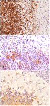
Mycoplasma bovis, the most important primary pathogen in the family Mycoplasmataceae, causes pneumonia, arthritis, otitis media, and mastitis in cattle. Histopathologic pulmonary changes associated with M. bovis infection have been characterized as suppurative-to-caseonecrotic bronchopneumonia; infection in other organs has been reported in only a few studies that examined caseonecrotic endocarditis and suppurative meningitis. Granulomatous lesions associated with M. bovis infection have been reported only rarely. We studied the granulomatous inflammation associated with M. bovis infection in several organs of 21 Japanese Black cattle. M. bovis was detected by isolation and loop-mediated isothermal amplification methods; other bacteria were detected using culture on 5% blood sheep agar and a MALDI-TOF MS Biotyper. Tissues were examined by histopathology and by immunohistochemistry (IHC) using anti-M. bovis, anti-Iba1, anti-iNOS, and anti-CD204 antibodies. All 21 cases, which included 2 cases of meningitis-meningoencephalitis, 8 cases of endocarditis, and 11 cases of bronchopneumonia, had caseonecrotic granulomatous inflammation associated with M. bovis infection. The IHC for macrophages revealed a predominance of iNOS-labeled (M1) macrophages in the inner layer of the caseonecrotic granulomas associated with meningitis-meningoencephalitis, endocarditis, and bronchopneumonia in Japanese Black cattle naturally infected with M. bovis.
Keywords: Mycoplasma bovis; caseous necrosis; cattle; classical activated macrophage; granulomatous inflammation.
Conflict of interest statement
Figures


Similar articles
-
Mycoplasma bovis May Travel Along the Eustachian Tube to Cause Meningitis in Japanese Black Cattle.J Comp Pathol. 2021 Oct;188:13-20. doi: 10.1016/j.jcpa.2021.08.001. Epub 2021 Sep 10. J Comp Pathol. 2021. PMID: 34686272
-
Naturally occurring Mycoplasma bovis-associated pneumonia and polyarthritis in feedlot beef calves.J Vet Diagn Invest. 2006 Jan;18(1):29-40. doi: 10.1177/104063870601800105. J Vet Diagn Invest. 2006. PMID: 16566255
-
Effects of inflammatory stimuli on the development of Mycoplasma bovis pneumonia in experimentally challenged calves.Vet Microbiol. 2024 Oct;297:110203. doi: 10.1016/j.vetmic.2024.110203. Epub 2024 Jul 30. Vet Microbiol. 2024. PMID: 39089141
-
Mycoplasma bovis in respiratory disease of feedlot cattle.Vet Clin North Am Food Anim Pract. 2010 Jul;26(2):365-79. doi: 10.1016/j.cvfa.2010.03.003. Epub 2010 May 14. Vet Clin North Am Food Anim Pract. 2010. PMID: 20619190 Review.
-
Is Mycoplasma bovis a missing component of the bovine respiratory disease complex in Australia?Aust Vet J. 2014 Jun;92(6):185-91. doi: 10.1111/avj.12184. Aust Vet J. 2014. PMID: 24862996 Review.
Cited by
-
An overview of the detection of bovine respiratory disease complex pathogens using immunohistochemistry: emerging trends and opportunities.J Vet Diagn Invest. 2024 Jan;36(1):12-23. doi: 10.1177/10406387231210489. Epub 2023 Nov 20. J Vet Diagn Invest. 2024. PMID: 37982437 Free PMC article. Review.
-
The Identification of a Novel Nucleomodulin MbovP467 of Mycoplasmopsis bovis and Its Potential Contribution in Pathogenesis.Cells. 2024 Mar 29;13(7):604. doi: 10.3390/cells13070604. Cells. 2024. PMID: 38607043 Free PMC article.
-
Influence of Paratuberculosis Vaccination on the Local Immune Response in Experimentally Infected Calves: An Immunohistochemical Analysis.Animals (Basel). 2025 Jun 22;15(13):1841. doi: 10.3390/ani15131841. Animals (Basel). 2025. PMID: 40646740 Free PMC article.
-
Intracranial inflammatory polyp with cerebellopontine compression and leptomeningitis secondary to chronic otitis in a red kangaroo.J Vet Diagn Invest. 2023 Nov;35(6):806-809. doi: 10.1177/10406387231195848. Epub 2023 Aug 24. J Vet Diagn Invest. 2023. PMID: 37615172 Free PMC article.
-
Mycoplasma bovis mastitis in dairy cattle.Front Vet Sci. 2024 Mar 6;11:1322267. doi: 10.3389/fvets.2024.1322267. eCollection 2024. Front Vet Sci. 2024. PMID: 38515536 Free PMC article. Review.
References
-
- Ackermann MR. Inflammation and healing. In: Zachary JF, ed. Pathologic Basis of Veterinary Disease. 6th ed. Elsevier, 2017:108–117.
-
- Ayling R, et al.. Mycoplasma bovis isolated from brain tissue of calves. Vet Rec 2005;156:391–392. - PubMed
-
- Caswell JL, Archambault M. Mycoplasma bovis pneumonia in cattle. Anim Health Res Rev 2007;8:161–186. - PubMed
-
- Caswell JL, Williams KJ. Respiratory system. In: Maxie MG, ed. Jubb, Kennedy, and Palmer’s Pathology of Domestic Animals. 6th ed. Vol. 2. Elsevier, 2016:551–554.
MeSH terms
LinkOut - more resources
Full Text Sources
Medical

