Case Reports
. 2021 Nov 13:14:3161-3175.
doi: 10.2147/JMDH.S330094. eCollection 2021.

Evaluation of Tibia Bone Healing by Infrared Thermography: A Case Study

Affiliations
Case Reports

Evaluation of Tibia Bone Healing by Infrared Thermography: A Case Study

Wally Auf der Strasse et al. J Multidiscip Healthc. .

Abstract

Background: Thermal imaging has been used as a clinical follow-up technique in several medical specialties.

Purpose: The aim of this study was to investigate the feasibility of using medical thermography in the diagnosis and follow-up assessment of a severe orthopedic trauma that requires the use of an external circular fixator.

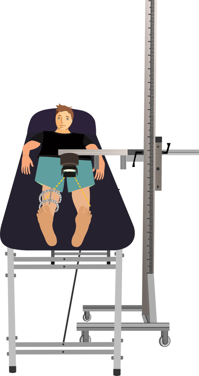
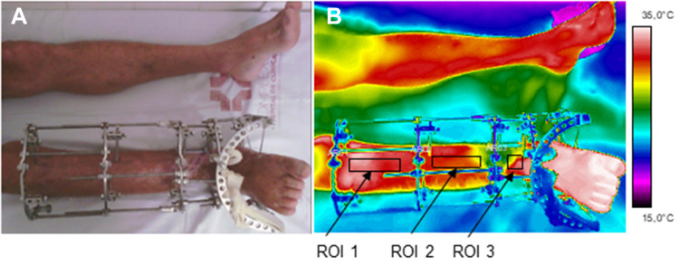
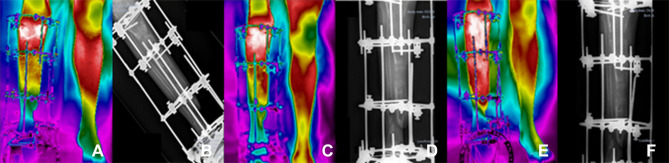
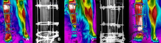
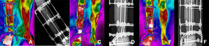
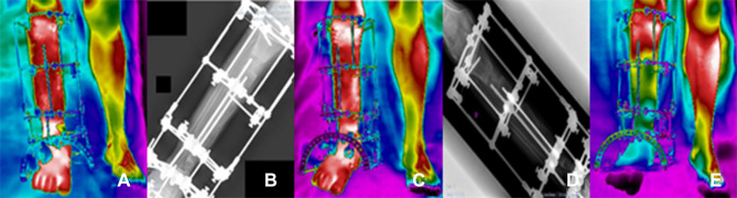
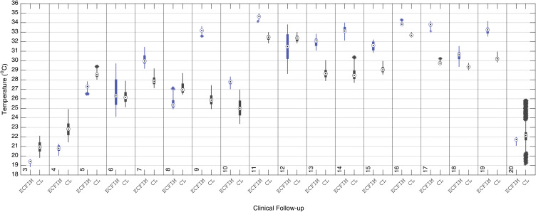
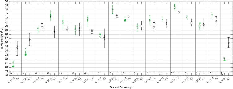
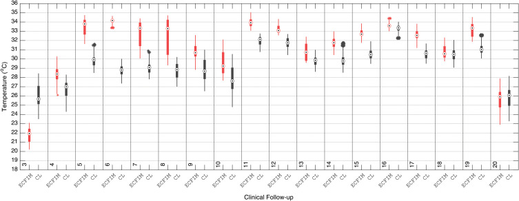
Patients and methods: Twenty clinical follow-ups of thermal imaging correlated with X-ray images were performed in a male volunteer, diagnosed with bone nonunion, during 11 months of treatment, in the hospital trauma and reconstruction department. Data were acquired in the regions of interest of the proximal tibia, diaphysis and distal, with a Flir T530 medical grade infrared camera from Flir Systems®, and the data processed by the Matlab® 2019 custom made software.

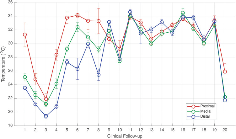
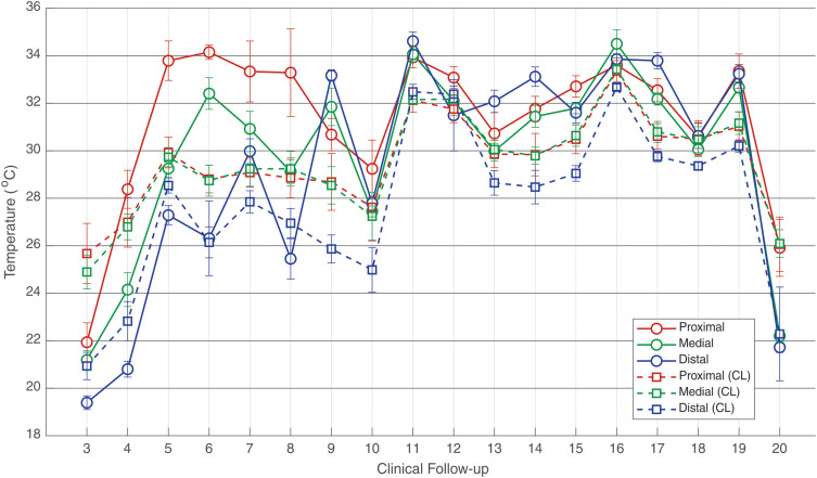
Results: Statistical analysis was performed by Wilcoxon signed-rank test. The results showed a median temperature of 22.2°C, and thus some periods of interruption in the healing process between the third and twentieth clinical follow-up, and a significant increase of the temperature to 34.6°C synchronous with a diagnosis of bone infection by the eleventh clinical follow-up. The thermal images acquired during the 20 clinical follow-ups allow a correlation with the data from the X-ray exams and also with the contralateral limb of the evaluated patient, showing thermal alterations greater than 0.3°C, which are significant of physiological abnormality.

Conclusion: The thermography exam can be a useful tool for applying on the follow-up of patients after trauma or bone fracture. The results showed important physiological data related to the vascularization necessary for bone repairing, being therefore a good indicator of the healing process. In addition, as infrared thermography does not use ionizing radiation, it can be used countlessly, in complement to the traditional X-ray exams that focus on anatomical data analysis.

Keywords: Ilizarov method; bone healing; infrared medical thermography; nonunion.


Conflict of interest statement

The authors report no conflicts of interest in this work.

Figures

Figure 1
Figure 1
Illustration schematic of the image acquisition setup.
Figure 2
Figure 2
Photograph of the evaluated limb (right leg) (A); Infrared image and defined ROIs (B). ROI1 –proximal tibia. ROI2 – tibia shaft (diaphyseal), ROI3 – distal tibia.
Figure 3
Figure 3
Clinical follow-ups 1 (A and B), 2 (C and D) and 3 (E and F); (A, C, E) infrared images and (B, D, F) X-ray.
Figure 4
Figure 4
Clinical follow-ups 4 (A and B), 5 (C and D) and 6 (E and F); (A, C, E) infrared images and (B, D, F) X-ray.
Figure 5
Figure 5
Clinical follow-ups 7 (A and B), 8 (C and D) and 9 (F), (A, C, F) infrared image and (B and D) X-ray.
Figure 6
Figure 6
Clinical follow-ups 12 (A and B), 14 (C and D) and 16 (E and F), (A, C, E) infrared image and (B, D, F) X-ray.
Figure 7
Figure 7
Clinical follow-ups 17 (A and B), 19 (C and D) and 20 (E), (A, C, E) infrared image and (B, D) X-ray.
Figure 8
Figure 8
Evolution of median temperature in the ROIs of the right leg (under treatment) in the 20 clinical follow-ups. The error bars correspond to one standard deviation.
Figure 9
Figure 9
Evolution of median temperature in the ROIs of the right leg (under treatment) in the 20 clinical segments using the ECFIM in comparison with the ROIs of the healthy left leg (CL). The error bars correspond to one standard deviation.
Figure 10
Figure 10
Boxplot of the median temperature of the ROI of the right proximal tibia with the use of the ECFIM, in the 20 follow-up sessions, compared with the ROI of the same portion of the healthy left leg (CL).
Figure 11
Figure 11
Boxplot of the median temperature of the ROI of the diaphysis portion of the right tibia using the ECFIM, in the 20 follow-up sessions, compared with the healthy left leg (CL).
Figure 12
Figure 12
Boxplot of the median temperature of the ROI of the right distal tibia portion (with ECFIM), in the following 20 clinicians, with the ROI of the same portion of the healthy left leg.

References

    1. Oliveira O, Martins SPR, Lima W, Gomes MM. The use of bone morphogenetic proteins (BMP) and pseudarthrosis, a literature review. Revista Brasileira De Ortopedia. 2017;52:124–140. doi:10.1016/j.rboe.2016.03.005 - DOI - PMC - PubMed
    1. Ulson O, Zamboni C, Durigan JR, et al. Treatment of femur pseudoarthrosis using wave plate: evaluation of consolidation and its relationship with graft type. Injury. 2021;52:S18–S22. doi:10.1016/j.injury.2021.01.045 - DOI - PubMed
    1. Ilizarov G, Deviatov A, Trokhova V. Surgical lengthening of the shortened lower extremities. Vestnik Khirurgii Imeni II Grekova. 1972;108(2):100–103. - PubMed
    1. Shortt NL, Keenan GF, Muir AY, Simpson A. The use of a nerve stimulator to allow safe placement of Ilizarov wires. Oper Orthop Traumatol. 2006;18(4):364–376. doi:10.1007/s00064-006-1177-2 - DOI - PubMed
    1. Peng J, Min L, Xiang Z, Huang F, Tu C, Zhang H. Ilizarov bone transport combined with antibiotic cement spacer for infected tibial nonunion. Int J Clin Exp Med. 2015;8(6):10058. doi:10.3390/ijerph18084217 - DOI - PMC - PubMed

Publication types

LinkOut - more resources