. 2021 Sep 16;49(5):439-453.
doi: 10.1080/12298093.2021.1965706. eCollection 2021.

Phallus chiangmaiensis sp. nov. and a Record of P. merulinus in Thailand

Affiliations

Phallus chiangmaiensis sp. nov. and a Record of P. merulinus in Thailand

Sujinda Sommai et al. Mycobiology. .

Abstract

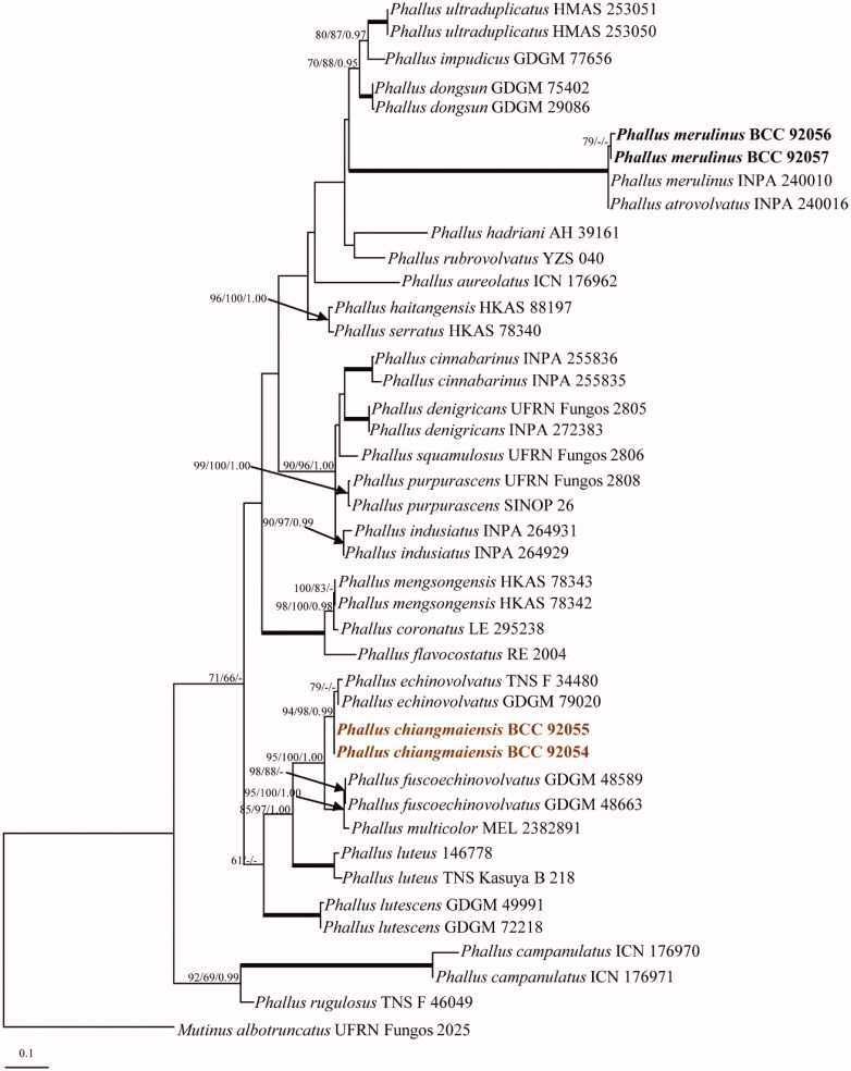
During the rainy season in Thailand, specimens of Phallus chiangmaiensis sp. nov. and P. merulinus were collected from Chiang Mai and Samut Sakhon Provinces, respectively. Molecular phylogenetic analyses based on sequences of the nuclear ribosomal large subunit (LSU), nuclear ribosomal 5.8S gene including the internal transcribed spacer regions 1 and 2 (ITS), and the protein-coding gene atp6 (mitochondrial adenosine triphosphate [ATP] synthase subunit 6) support the placement of the new species within Phallus. Phallus chiangmaiensis has a well-developed white indusium and campanulated caps with reticulate surfaces. It differs morphologically from the related species, as supported by the phylogenetic data. Phallus merulinus is reported here as a species that was re-encountered in Thailand. The descriptions of the species are accompanied by illustrations of macro- and micro- morphological features, and a discussion of the related taxa is presented.

Keywords: Stinkhorn fungus; phylogeny; taxonomy.


Conflict of interest statement

No potential conflict of interest was reported by the author(s).

Figures

Figure 1.
Figure 1.
Phylogenetic relationships of Phallus spp. inferred from ITS sequences. Numbers at the significant nodes represent ML bootstrap values/MP/Bayesian posterior probabilities, multiplied by 100; bold lines in the tree represent 100% bootstrap (BSMP, BSML) and 1.00 posterior probability (BPP).
Figure 2.
Figure 2.
Phylogenetic relationships of Phallus spp. from a combined ITS, LSU, and atp6 analyses. Numbers at the significant nodes represent ML bootstrap values/MP/Bayesian posterior probabilities, multiplied by 100; bold lines in the tree represent 100% bootstrap (BSMP, BSML) and 1.00 posterior probability (BPP).
Figure 3.
Figure 3.
Phallus chiangmaiensis (BBH 47825, holotype). (a) Mature basidiomata. (b) Reticulate cap. (c) Indusium. (d) Immature basidiomata (egg). (e) Pseudostipe and section of immature basidiomata. Scale bars: a = 50 mm, b–e = 10 mm.
Figure 4.
Figure 4.
Microscopic features of Phallus chiangmaiensis. (a,b) Cap cells and hyphae. (c) Cells of indusium. (d) Cells of pseudostipe. (e) Crystals in volva hyphae (arrowed). (f,g) Volva hyphae with clamp connections (arrowed). (h,i) Rhizomorph hyphae with clamp connections (arrowed). (j) Basidia with sterigmata and basidiospores. (k) Basidiospores. (l) Colony on PDA (surface and reverse plate). Scale bars: a, c–e = 20 µm, b, f–j = 10 µm, g, k = 5 µm, l = 10 mm.
Figure 5.
Figure 5.
Line drawing of Phallus chiangmaiensis. (a) Fruiting body. (b) Cells of indusium. (c,d) Cells of pseudostipe. (e) Volva hyphae. (f) Rhizomorph hyphae. (g) Basidia. (h) Basidiospores. Scale bars: a = 50 mm, b–d = 50 μm, e = 10 μm, f = 20 μm, g–h = 5 μm.
Figure 6.
Figure 6.
Phallus merulinus (BBH 47826). (a,b) Basidiomata. (c,d) Immature basidiomata (eggs) with rhizomorph (arrowed). (e) Indusium and pseudostipe. (f) Volva. Scale bars: a–b = 50 mm, c–f = 20 mm.
Figure 7.
Figure 7.
Microscopic features of Phallus merulinus. (a–e) Cap cells and hyphae. (f) Cells of indusium. (g) Cells of pseudostipe. (h,i) Volva hyphae. (j,k) Rhizomorph hyphae. (l) Basidiospores. (m) Colony on PDA (surface and reverse plate). Scale bars: a–b, f–g = 20 µm, c–e, h–l = 5 µm, m = 20 mm.
Figure 8.
Figure 8.
Line drawing of Phallus merulinus. (a) Fruiting body. (b,c) Cap cells and hyphae. (d) Cells of indusium. (e,f). Cells of pseudostipe. (g) Volva hyphae. (h) Rhizomorph hyphae. (i) Basidiospores. Scale bars: a = 20 mm, b, g = 10 μm, c, h, i = 5 μm, d = 20 μm, e–f = 50 μm.

References

    1. Arora D. Mushrooms demystified: a comprehensive guide to the fleshy fungi. Berkeley: Ten Speed Press; 1986. p. 1056.
    1. Kreisel H. A preliminary survey of the genus Phallus sensu lato. Czech Mycol. 1996;48(4):273–281.
    1. Liu B, Fan L, Li JZ, et al. . Flora fungorum sinicorum. Vol. 23. Beijing: Science Press; 2005. p. 137–171.
    1. Dai YC, Yang ZL.. A revised checklist of medicinal fungi in China. Mycosystema. 2008;27:801–824.
    1. Dai YC, Zhou LW, Yang ZL, et al. . A revised checklist of edible fungi in China. Mycosystema. 2010;29:1–21.