. 2021 Nov 4:15:734158.
doi: 10.3389/fnins.2021.734158. eCollection 2021.

Hippocampal Function Is Impaired by a Short-Term High-Fat Diet in Mice: Increased Blood-Brain Barrier Permeability and Neuroinflammation as Triggering Events

Affiliations

Hippocampal Function Is Impaired by a Short-Term High-Fat Diet in Mice: Increased Blood-Brain Barrier Permeability and Neuroinflammation as Triggering Events

Gabriela Cristina de Paula et al. Front Neurosci. .

Abstract

Worldwide, and especially in Western civilizations, most of the staple diets contain high amounts of fat and refined carbohydrates, leading to an increasing number of obese individuals. In addition to inducing metabolic disorders, energy dense food intake has been suggested to impair brain functions such as cognition and mood control. Here we demonstrate an impaired memory function already 3 days after the start of a high-fat diet (HFD) exposure, and depressive-like behavior, in the tail suspension test, after 5 days. These changes were followed by reduced synaptic density, changes in mitochondrial function and astrocyte activation in the hippocampus. Preceding or coinciding with the behavioral changes, we found an induction of the proinflammatory cytokines TNF-α and IL-6 and an increased permeability of the blood-brain barrier (BBB), in the hippocampus. Finally, in mice treated with a TNF-α inhibitor, the behavioral and BBB alterations caused by HFD-feeding were mitigated suggesting that inflammatory signaling was critical for the changes. In summary, our findings suggest that HFD rapidly triggers hippocampal dysfunction associated with BBB disruption and neuroinflammation, promoting a progressive breakdown of synaptic and metabolic function. In addition to elucidating the link between diet and cognitive function, our results might be relevant for the comprehension of the neurodegenerative process.

Keywords: bioenergetics; blood–brain barrier; cognition; depression; high fat diet; memory; mitochondria; neuroinflammation.


Conflict of interest statement

The authors declare that the research was conducted in the absence of any commercial or financial relationships that could be construed as a potential conflict of interest.

Figures

FIGURE 1
FIGURE 1
Hippocampal abnormalities of mice fed a high-fat diet. (A) First time-course experimental design. (B) Novel object recognition (NOR) and (C) tail suspension tests (TSTs) were performed in mice fed SD or HFD for 1, 2, and 4 weeks. Cognitive performance of SD and HFD-treated mice was accessed by NOR (n = 7–8/group; one-sample t-test with hypothetical value of 50%). Time of immobility in TST was recorded in order to evaluate depressive-like behavior (n = 7–8; unpaired t-test for 1, 2, and 4 weeks of diet, respectively). (D) Sodium fluorescein assay to evaluate BBB permeability on hippocampus (n = 5–7; t-test for 1, 2, and 4 weeks of diet). Values are mean ± SEM. +Means higher than the hypothetical value of 50%, *p < 0.05 SD vs. HFD, +discrimination score above 50%.
FIGURE 2
FIGURE 2
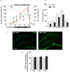
Hippocampal alterations followed by 4 weeks of HFD. (A) Representative experiment in SUIT protocol. (B) Respirometric measurements by oxygen flux using PM as initial substrates (leak), followed by ADP (CIp), S (Oxphos), FCCP titrations (ETS), and finally inhibited by Rot (CIIp; F = 2.077) and Ant A in the hippocampus of mice submitted to 4 weeks of SD or HFD (n = 5–6). (C) Representative images of GFAP immunolabeling in the hippocampus of 4 weeks SD and HFD-fed mice (scale bar = 150 μm) along with (D) quantification of GFAP immunoreactivity (n = 4). Values are mean ± SEM. Unpaired t-test performed. *p < 0.05, **p < 0.01 SD vs. HFD.
FIGURE 3
FIGURE 3
Evaluation of the high-fat diet effects in an early time-course. Assessment of the onset changes in (A) cognition (n = 6–8/group; one-sample t-test with hypothetical value of 50%), (B) depressive-like behavior (n = 6–8), (C) BBB leakage in hippocampus of mice fed HFD from 1 up to 6 days (n = 4), as well as proinflammatory cytokines represented by (D) TNF-α, (E) IL-6, and (F) IL-1β (n = 4). One-way ANOVA followed by Dunnett post hoc test performed for (B–F). *p < 0.05, ***p < 0.001 SD vs. HFD. Values are mean ± SEM. +p < 0.05 vs. chance levels (50% of a new object investigation in test trial).
FIGURE 4
FIGURE 4
Decrease of synaptic density after 7 days of HFD. (A) Immunofluorescence analysis for the expression and localization of the pre-synaptic terminals immunolabeled for pre-synaptic marker synaptophysin (SYP) in hippocampal CA1 region of mice submitted to SD (0), 3 or 7 days of HFD (n = 4–5, one-way ANOVA followed by Dunnett post hoc test). SYP represented in green and DAPI represented in blue. Scale bar = 50 μm (B) quantitative analysis for the SYP immunoreactivity. Data are shown as mean ± SEM. *p < 0.05 SD vs. HFD.
FIGURE 5
FIGURE 5
Anti-inflammatory effect on CNS alterations induced by high-fat diet. (A) Experimental design of mice fed either SD or HFD for 7 days and treated with saline or infliximab (10 mg/kg). (B) Cognitive performance of treated mice (n = 7–8/group; one-sample t-tests). (C) Time of immobility in TST (n = 7–8; two-way ANOVA followed by Duncan post hoc test). (D) Hippocampal sodium fluorescein assay (n = 4/group; two-way ANOVA followed by Duncan post hoc test). Values are mean ± SEM. +p < 0.05 vs. chance levels (50% of a new object investigation in test trial). **p < 0.01 compared with mice fed with SD and &p < 0.05 compared HFD mice treated with vehicle. *p < 0.05, **p < 0.01, ***p < 0.001 SD vs. HFD.

Similar articles

Cited by

References

    1. Abildgaard A., Solskov L., Volke V., Harvey B. H., Lund S., Wegener G. (2011). A high-fat diet exacerbates depressive-like behavior in the flinders sensitive line (FSL) rat, a genetic model of depression. Psychoneuroendocrinology 36 623–633. 10.1016/j.psyneuen.2010.09.004 - DOI - PubMed
    1. Araújo E. P., De Souza C. T., Ueno M., Cintra D. E., Bertolo M. B., Carvalheira J. B., et al. (2007). Infliximab restores glucose homeostasis in an animal model of diet-induced obesity and diabetes. Endocrinology 148 5991–5997. 10.1210/en.2007-0132 - DOI - PubMed
    1. Arroyo-Johnson C., Mincey K. D. (2016). Obesity epidemiology worldwide. Gastroenterol. Clin. North Am. 45 571–579. 10.1016/j.gtc.2016.07.012 - DOI - PMC - PubMed
    1. Asfaw A. (2011). Does consumption of processed foods explain disparities in the body weight of individuals? The case of guatemala. Health Econ. 20 184–195. 10.1002/hec.1579 - DOI - PubMed
    1. Attuquayefio T. N., Stevenson R. J., Oaten M. J., Francis H. M. (2017). A four-day Western-style dietary intervention causes reductions in hippocampal-dependent learning and memory and interoceptive sensitivity. PLoS One 12:e0172645. 10.1371/journal.pone.0172645 - DOI - PMC - PubMed