

Psychological Stress as a Risk Factor for Accelerated Cellular Aging and Cognitive Decline: The Involvement of Microglia-Neuron Crosstalk
- PMID: 34803607
- PMCID: PMC8599581
- DOI: 10.3389/fnmol.2021.749737
Psychological Stress as a Risk Factor for Accelerated Cellular Aging and Cognitive Decline: The Involvement of Microglia-Neuron Crosstalk
Abstract
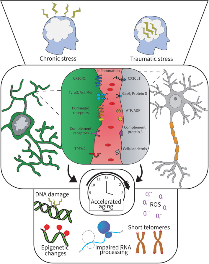
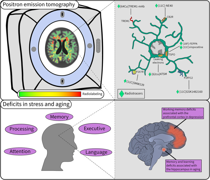
The relationship between the central nervous system (CNS) and microglia is lifelong. Microglia originate in the embryonic yolk sac during development and populate the CNS before the blood-brain barrier forms. In the CNS, they constitute a self-renewing population. Although they represent up to 10% of all brain cells, we are only beginning to understand how much brain homeostasis relies on their physiological functions. Often compared to a double-edged sword, microglia hold the potential to exert neuroprotective roles that can also exacerbate neurodegeneration once compromised. Microglia can promote synaptic growth in addition to eliminating synapses that are less active. Synaptic loss, which is considered one of the best pathological correlates of cognitive decline, is a distinctive feature of major depressive disorder (MDD) and cognitive aging. Long-term psychological stress accelerates cellular aging and predisposes to various diseases, including MDD, and cognitive decline. Among the underlying mechanisms, stress-induced neuroinflammation alters microglial interactions with the surrounding parenchymal cells and exacerbates oxidative burden and cellular damage, hence inducing changes in microglia and neurons typical of cognitive aging. Focusing on microglial interactions with neurons and their synapses, this review discusses the disrupted communication between these cells, notably involving fractalkine signaling and the triggering receptor expressed on myeloid cells (TREM). Overall, chronic stress emerges as a key player in cellular aging by altering the microglial sensome, notably via fractalkine signaling deficiency. To study cellular aging, novel positron emission tomography radiotracers for TREM and the purinergic family of receptors show interest for human study.
Keywords: chronic psychological stress; cognitive aging; major depressive disorder; microglia; neuron; oxidative stress; synapse.
Copyright © 2021 Carrier, Šimončičová, St-Pierre, McKee and Tremblay.
Conflict of interest statement
The authors declare that the research was conducted in the absence of any commercial or financial relationships that could be construed as a potential conflict of interest.
Figures
References
Publication types
LinkOut - more resources
Full Text Sources
Research Materials