. 2021 Nov 4:12:724417.
doi: 10.3389/fneur.2021.724417. eCollection 2021.

The Onset of Interictal Spike-Related Ripples Facilitates Detection of the Epileptogenic Zone

Affiliations

The Onset of Interictal Spike-Related Ripples Facilitates Detection of the Epileptogenic Zone

Yurika Numata-Uematsu et al. Front Neurol. .

Abstract

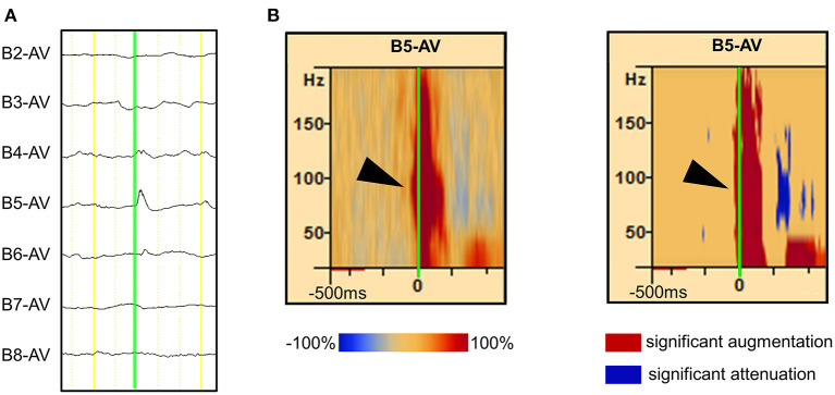
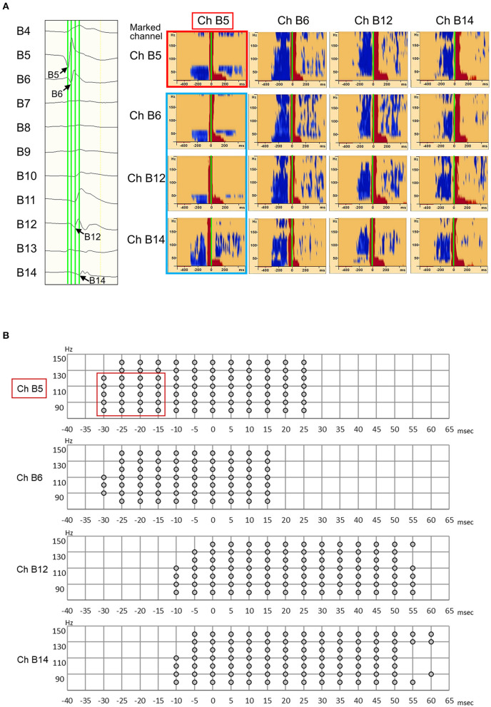
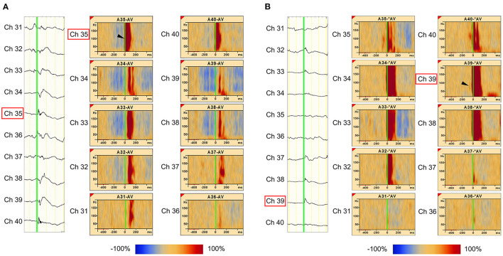
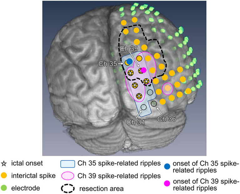
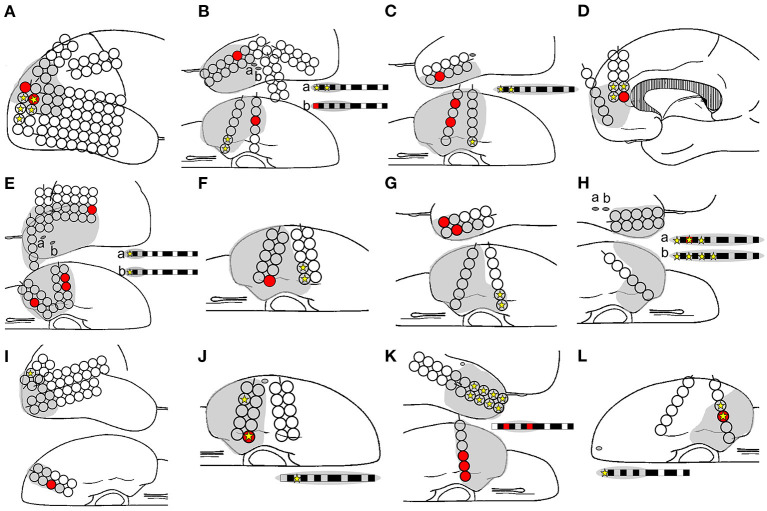
Objective: Accurate estimation of the epileptogenic zone (EZ) is essential for favorable outcomes in epilepsy surgery. Conventional ictal electrocorticography (ECoG) onset is generally used to detect the EZ but is insufficient in achieving seizure-free outcomes. By contrast, high-frequency oscillations (HFOs) could be useful markers of the EZ. Hence, we aimed to detect the EZ using interictal spikes and investigated whether the onset area of interictal spike-related HFOs was within the EZ. Methods: The EZ is considered to be included in the resection area among patients with seizure-free outcomes after surgery. Using a complex demodulation technique, we developed a method to determine the onset channels of interictal spike-related ripples (HFOs of 80-200 Hz) and investigated whether they are within the resection area. Results: We retrospectively examined 12 serial patients who achieved seizure-free status after focal resection surgery. Using the method that we developed, we determined the onset channels of interictal spike-related ripples and found that for all 12 patients, they were among the resection channels. The onset frequencies of ripples were in the range of 80-150 Hz. However, the ictal onset channels (evaluated based on ictal ECoG patterns) and ripple onset channels coincided in only 3 of 12 patients. Conclusions: Determining the onset area of interictal spike-related ripples could facilitate EZ estimation. This simple method that utilizes interictal ECoG may aid in preoperative evaluation and improve epilepsy surgery outcomes.

Keywords: epilepsy; epileptogenic zone; interictal; onset; spike-related ripples; surgery.


Conflict of interest statement

The authors declare that the research was conducted in the absence of any commercial or financial relationships that could be construed as a potential conflict of interest.

Figures

Figure 1
Figure 1
Detection of spike-related high-frequency oscillations (HFOs) using the BESA® software. (A) The rising edge of the negative spike is marked (green line) in the channel of interest [channel (Ch) B5 is used as an example in this figure]. More than 30 spikes were marked in the same channel. (B, left) The average HFO augmentation associated with the spikes was analyzed using BESA®. Within the square, the “percentage augmentation” of the Ch B5 spike-related HFOs relative to the reference (300–500 ms prior to the spike, red bar) is shown, ranging from −100% (blue) to +100% (red). The earliest HFO augmentation was observed between 80 and 150 Hz (arrowhead). (B, right) The blue color indicates the significant attenuation and the red color indicates the significant augmentation of amplitude in a given time–frequency bin relative to the reference period. The analysis was done using studentized bootstrap statistics. The corrected α level was set to 0.05.
Figure 2
Figure 2
Illustration showing high-frequency oscillation (HFO) onset in Ch B5 for all marked channels (B5, B6, B12, and B14). (A, left) Interictal electrocorticography (ECoG) traces are shown, along with interictal spike propagation. In the enlarged ECoG trace, marks (green line) are attached to the rising edge of the Ch B5, Ch B6, Ch B12, and Ch B14 spikes. (A, right) The Ch B5, Ch B6, Ch B12, and Ch B14 spike-related HFOs are shown at the top, second from top, third from top, and at the bottom, respectively. The blue color indicates significant HFO attenuation and the red color indicates significant HFO augmentation in a given time–frequency bin relative to the reference period. At the top, the HFO onset of Ch B5 spike-related HFOs were observed as Ch B5 (red frame). In the other three channels, spike-related HFOs also have their onset in Ch B5 (blue frame). Thus, HFO onset can be accurately identified as Ch B5 by marking any channel (Ch B5, Ch B6, Ch B12, and Ch B14) over which the spikes spread. (B) Spike-related ripple onset in Ch B5. In our novel method, we defined significant HFO amplitude modulation as at least 40 Hz in width and 20 ms in duration. Spike-related ripple onset in Ch B5, with a latency of −30 ms, from 80 to 130 Hz (red frame). The spike-related ripples of Ch B6, Ch B12, and Ch B14 had a slightly longer latency than those of Ch B5.
Figure 3
Figure 3
Marked interictal spikes and spike-related ripples in Patient 1. Green lines on the rising edge of channel A35 (Ch 35) [negative spike (A, left)] and channel A39 (Ch 39) [negative spikes (B, left)] are shown. The time–frequency plots in the square show the augmentation of ripples related to Ch 35 spikes (A, right) and Ch 39 spikes (B, right). Augmentation of ripples ranges from −100% (blue) to 100% (red) relative to the reference period (−500 to −300 ms). In the middle column of Ch 35 spike-related ripples (A), the red bands from Ch 35 to Ch 31 show a progressively longer latency, which indicates propagation of ripples across these channels. The shortest latency of the Ch 35 spikes-related ripple augmentation channel (ripple onset) was visually estimated as Ch 35 (A, arrowhead), and the Ch 39 spikes-related ripple augmentation channel (ripple onset) was estimated as Ch 39 (B, arrowhead).
Figure 4
Figure 4
Ripple onset in Ch 35 and Ch 39, and other findings in Patient 1. Spike-related ripples of Ch 35 and Ch 39 (light pink and light blue, respectively), ripple onset in Ch 35 and Ch 39 (filled pink circles and filled blue circles, respectively), ictal onset (star), interictal spike (filled yellow circle), placed electrode (filled green circle), and the resection margin (black dotted line) are shown. The spike-related ripples of each channel propagated around the ictal onset area and had a narrower distribution than the spikes. Ch 35 is not estimated as the ictal onset, but rather as the ripple onset. Ch 39 was estimated as both ictal and ripple onset.
Figure 5
Figure 5
Relationships among ripple onset, ictal onset, and the resection area. The ripple onset electrode (filled red circle and box), ictal onset electrode (filled yellow star), and resection area (gray area) are shown. Letters (A–L) in the schema represent Patients 1–12. The ripple onset channel was within the resection area in all the patients. The ripples and ictal channels were identical for only three patients: Patients 8 (H), 10 (J), and 12 (L).

References

    1. Wendling F, Bartolomei F, Bellanger JJ, Bourien J, Chauvel P. Epileptic fast intracerebral EEG activity: evidence for spatial decorrelation at seizure onset. Brain. (2003) 126:1449–59. 10.1093/brain/awg144 - DOI - PMC - PubMed
    1. Grinenko O, Li J, Mosher JC, Wang IZ, Bulacio JC, Gonzalez-Martinez J, et al. . A fingerprint of the epileptogenic zone in human epilepsies. Brain. (2018) 141:117–31. 10.1093/brain/awx306 - DOI - PMC - PubMed
    1. Gnatkovsky V, de Curtis M, Pastori C, Cardinale F, Lo Russo G, Mai R, et al. . Biomarkers of epileptogenic zone defined by quantified stereo-EEG analysis. Epilepsia. (2014) 55:296–305. 10.1111/epi.12507 - DOI - PubMed
    1. Sato Y, Wong SM, Iimura Y, Ochi A, Doesburg SM, Otsubo H. Spatiotemporal changes in regularity of gamma oscillations contribute to focal ictogenesis. Sci Rep. (2017) 7:9362. 10.1038/s41598-017-09931-6 - DOI - PMC - PubMed
    1. Akiyama T, McCoy B, Go CY, Ochi A, Elliott IM, Akiyama M, et al. . Focal resection of fast ripples on extraoperative intracranial EEG improves seizure outcome in pediatric epilepsy. Epilepsia. (2011) 52:1802–11. 10.1111/j.1528-1167.2011.03199.x - DOI - PubMed

LinkOut - more resources