. 2021 Nov 5:12:716314.
doi: 10.3389/fimmu.2021.716314. eCollection 2021.

Splenic Transcriptional Responses in Severe Visceral Leishmaniasis: Impaired Leukocyte Chemotaxis and Cell Cycle Arrest

Affiliations

Splenic Transcriptional Responses in Severe Visceral Leishmaniasis: Impaired Leukocyte Chemotaxis and Cell Cycle Arrest

Caroline Vilas Boas de Melo et al. Front Immunol. .

Abstract

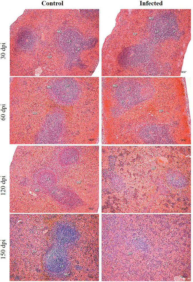
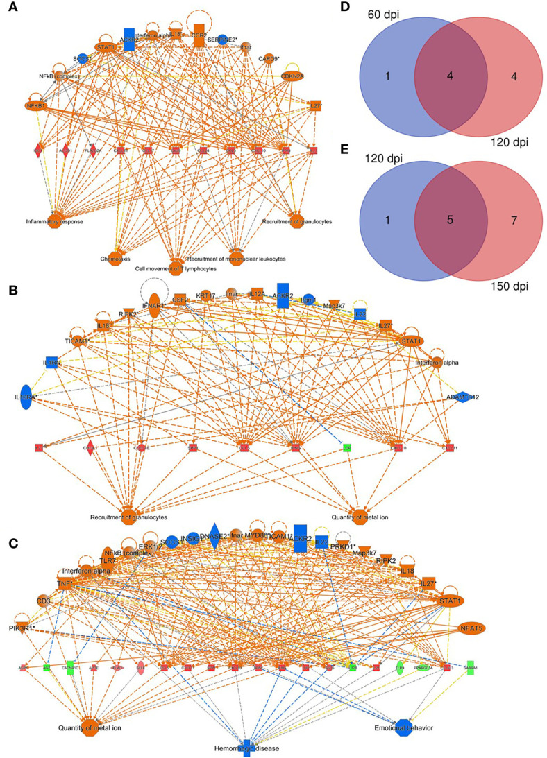
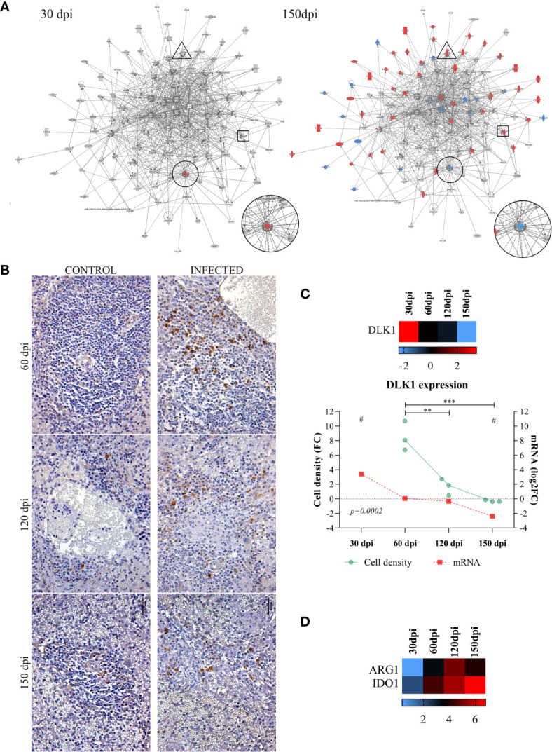
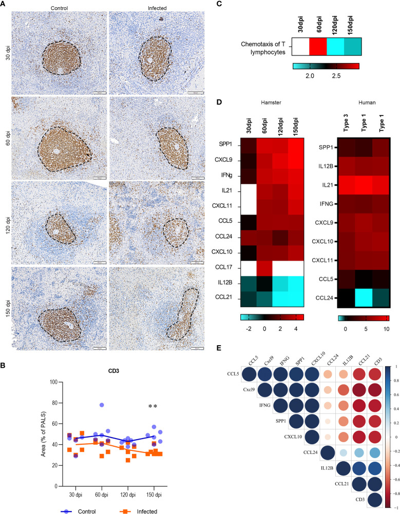
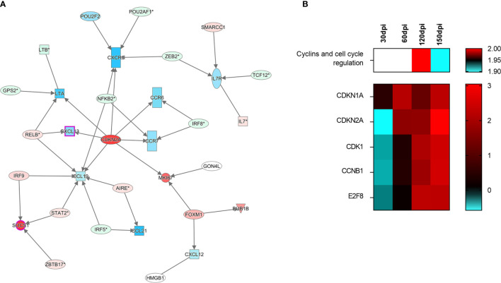
Structural changes in the spleen have been reported in several infectious diseases. In visceral leishmaniasis (VL), a severe parasitic disease caused by Leishmania spp., the loss of white pulp accompanies a severe clinical presentation. Hamster model reproduces aspects of human VL progression. In the early stages, a transcriptomic signature of leukocyte recruitment was associated with white pulp hyperplasia. Subsequently, impaired leukocyte chemotaxis with loss of T lymphocytes in the periarteriolar lymphoid sheath occurred. This differential gene expression was subsequently corroborated by transcriptomic profiling of spleens in severe human VL. At the latest stage, spleen disorganization was associated with increasing clinical signs of VL. White pulp disruption was accompanied by decreased DLK1 expression. The expression of CXCL13, CCR5, CCL19, CCR6, CCR7 and LTA decreased, likely regulated by CDKN2A overexpression. Our findings enlighten a pathway implying cell cycle arrest and decreased gene expression involved in spleen organization.

Keywords: hamster; spleen disorganization; spleen pathology; transcriptomic (RNA-Seq); visceral leishmanaisis; white pulp remodeling.


Conflict of interest statement

The authors declare that the research was conducted in the absence of any commercial or financial relationships that could be construed as a potential conflict of interest.

Figures

Figure 1
Figure 1
Histological analysis of hamster spleens. Representative photos of hamster spleens in control and infected groups (30-, 60-, 120- and 150 days post infection—dpi). H&E staining, 40x magnification, bar = 100 micrometers. Type I spleens are seen in all photomicrographs, except for infected at 120 dpi (type 2) and infected at 150 dpi (type 3). RP, red pulp; MZ, marginal zone; LF, lymphoid follicle; GC, germinal center; P,periarteriolar lymphoid sheath.
Figure 2
Figure 2
Regulatory effectors of splenic response to Leishmania infantum infection. (A–C) Network of transcripts associated with inflammatory response processes, chemotaxis, T lymphocyte movement, recruitment of mononuclear leukocytes and recruitment of granulocytes, quantity of metal ion, hemorrhagic disease and behavior in hamsters at 60 (A), 120 (B) and 150 dpi (C). Patterns of expression: green = low; red = high. Secondary interactions of transcripts and transcript-associated processes: orange= predicted activation; blue= predicted inhibition; dotted lines= indirect interaction; continuous lines= direct interaction; gray lines= no predicted effect; yellow lines= inconsistent findings. Venn diagrams depict number of overlapping transcripts expression at 60 and 120 dpi (D) and at 120 and 150 dpi (E).
Figure 3
Figure 3
Common differentially expressed transcripts at different stages of Leishmania infantum infection. (A) Network of overlapping DE genes at 30 and 150 dpi. Pattern of expression in networks: blue = low; red = high; gray = no differential expression. DLK1 is indicated in circles, IDO1 in rectangles and ARG1 in triangle forms. (B) Immunohistochemistry of DLK1 protein (brown staining) in the spleen of control and infected hamsters at 60, 120 and 150 dpi, 400x magnification. Bars= 40µm. (C) Scatter plots of morphometric estimates of DLK1 protein expression from immunohistochemistry staining in the spleens of infected hamsters at 60, 120 and 150 dpi (green line and dots). Cell density per area values were expressed in fold change (FC) from control hamsters. Expression of DLK1 transcript evidenced by RNA-sequencing at 30, 60, 120 and 150 dpi (mRNA, red line and squares), and in heatmap represented by log2FC. Lines represent the mean of the values. **= statistical difference of cell density FC between 60 dpi and 120 (p=0.001) and ***= statistical difference of cell density FC between 150 dpi (p=0.0005), ANOVA, Tukey’s multiple comparison test. #= FDR for statistical significance of DLK1 transcript expression at 30 and 150 dpi (padj<0.0001). Test for linear trend was performed to cell density FC between time points, with statistical significance for linear reduction at each timepoint (p=0.0002). (D) Expression of ARG1 and IDO1 at 30, 60, 120 and 150 dpi represented in log2FC in heatmap. Pattern of expression in heatmap: blue = low; red = high; black = no differential expression.
Figure 4
Figure 4
Transcriptomic and tissue profiling of T cells in spleens of Leishmania infantum-infected hamsters. (A) Immunohistochemistry of splenic CD3 (brown staining) in PALS area (dotted line) of control and infected hamsters at 30, 60, 120 and 150 dpi, 200x magnification. Bars= 100µm. (B) Scatter plots of morphometric estimates of CD3 expression in the spleens of control (blue) and infected (orange) hamsters at 30, 60, 120 and 150 dpi. Values were expressed in percentages of positive staining for CD3 within the PALS area. Lines represent the median of the values. **= statistical difference between control and infected groups at 150 dpi, p=0.004, Kruskal-Wallis test. (C) Heatmap representing predicted function of chemotaxis of T lymphocytes in the dataset (log2FC). (D) Set of transcripts that jointly predict the function of T lymphocyte chemotaxis in experimental (30, 60, 120 and 150 dpi) and human VL. Spleens from humans were organized (type 1) or disorganized (type 3). Patterns of expression: blue = low; red = high; blank = no detection. (E) Correlation matrix between transcripts that jointly predict the function of T lymphocyte chemotaxis (SPP1, CXCL9, IFN-γ, CCL5, CCL24, CXCL10, CCL21 and IL12B – ) and CD3 expression in the spleen of infected hamsters at 30, 60, 120 and 150 dpi. Values were corrected by Log2 in comparison to respective control groups. Blue= positive correlations; Red= negative correlations. Color intensity and circle size are proportional to the correlation coefficient (r) of Pearson method.
Figure 5
Figure 5
Signaling of cell-cycle arrest in the spleen of hamsters infected with Leishmania infantum. (A) Interaction network of transcripts associated with splenic white pulp disorganization at 150 days post-infection. Blue and red shading indicates differing grades of expression. CDKN2A can be found at the center of the network. (B) Canonical pathway of cyclins and cell cycle regulation at different stages of infection and set of transcripts involved in signaling. Patterns of expression: blue = low; red = high; blank = no differential expression.

Similar articles

Cited by

References

    1. Melby PC, Chandrasekar B, Zhao W, Coe JE. The Hamster as a Model of Human Visceral Leishmaniasis: Progressive Disease and Impaired Generation of Nitric Oxide in the Face of a Prominent Th1-Like Cytokine Response. J Immunol (2001) 166:1912–20. doi: 10.4049/jimmunol.166.3.1912 - DOI - PubMed
    1. Moreira N das D, Vitoriano-Souza J, Roatt BM, Vieira PM de A, Ker HG, Cardoso JM de O, et al. . Parasite Burden in Hamsters Infected With Two Different Strains of Leishmania (Leishmania) Infantum: “Leishman Donovan Units” Versus Real-Time PCR. PloS One (2012) 7:e47907. doi: 10.1371/journal.pone.0047907 - DOI - PMC - PubMed
    1. Hermida M d’El-R, de Melo CVB, Lima IDS, Oliveira GGdeS, Dos-Santos WLC. Histological Disorganization of Spleen Compartments and Severe Visceral Leishmaniasis. Front Cell infection Microbiol (2018) 8:394. doi: 10.3389/fcimb.2018.00394 - DOI - PMC - PubMed
    1. Lage DP, Ludolf F, Silveira PC, MacHado AS, Ramos FF, Dias DS, et al. . Screening Diagnostic Candidates From Leishmania Infantum Proteins for Human Visceral Leishmaniasis Using an Immunoproteomics Approach. Parasitology (2019)146:1467–76. doi: 10.1017/S0031182019000714 - DOI - PubMed
    1. Lima IS, Silva JS, Almeida VA, Leal FG, Souza PAN, Larangeira DF, et al. . Severe Clinical Presentation of Visceral Leishmaniasis in Naturally Infected Dogs With Disruption of the Splenic White Pulp. PloS One (2014) 9:e87742. doi: 10.1371/journal.pone.0087742 - DOI - PMC - PubMed

Publication types

MeSH terms