. 2021 Nov 10:2021:7037786.
doi: 10.1155/2021/7037786. eCollection 2021.

Targeting the Ang2/Tie2 Axis with Tanshinone IIA Elicits Vascular Normalization in Ischemic Injury and Colon Cancer

Affiliations

Targeting the Ang2/Tie2 Axis with Tanshinone IIA Elicits Vascular Normalization in Ischemic Injury and Colon Cancer

Wei Zou et al. Oxid Med Cell Longev. .

Abstract

Pathological angiogenesis, as exhibited by aberrant vascular structure and function, has been well deemed to be a hallmark of cancer and various ischemic diseases. Therefore, strategies to normalize vasculature are of potential therapeutic interest in these diseases. Recently, identifying bioactive compounds from medicinal plant extracts to reverse abnormal vasculature has been gaining increasing attention. Tanshinone IIA (Tan IIA), an active component of Salvia miltiorrhiza, has been shown to play significant roles in improving blood circulation and delaying tumor progression. However, the underlying mechanisms responsible for the therapeutic effects of Tan IIA are not fully understood. Herein, we established animal models of HT-29 human colon cancer xenograft and hind limb ischemia to investigate the role of Tan IIA in regulating abnormal vasculature. Interestingly, our results demonstrated that Tan IIA could significantly promote the blood flow, alleviate the hypoxia, improve the muscle quality, and ameliorate the pathological damage after ischemic insult. Meanwhile, we also revealed that Tan IIA promoted the integrity of vascular structure, reduced vascular leakage, and attenuated the hypoxia in HT-29 tumors. Moreover, the circulating angiopoietin 2 (Ang2), which is extremely high in these two pathological states, was substantially depleted in the presence of Tan IIA. Also, the activation of Tie2 was potentiated by Tan IIA, resulting in decreased vascular permeability and elevated vascular integrity. Mechanistically, we uncovered that Tan IIA maintained vascular stability by targeting the Ang2-Tie2-AKT-MLCK cascade. Collectively, our data suggest that Tan IIA normalizes vessels in tumors and ischemic injury via regulating the Ang2/Tie2 signaling pathway.


Conflict of interest statement

The authors declared no conflict of interest.

Figures

Figure 1
Figure 1
Tan IIA improved blood perfusion recovery in the ischemic hind limbs. (a) Schematic diagram for depicting the establishment of the combined mouse model of HT-29 xenograft and hind limb ischemia, including the schedule of Tan IIA treatments. (b) Representative images of LDPI of the ischemic hind limbs in mice treated with 0.3% CMC-Na or Tan IIA at the indicated time points. (c) Hindlimb blood flow expressed as a percentage of ischemic limb blood flow over nonischemic hindlimb blood flow measured at the indicated time points (n = 6). (d) Morphology assessment of ischemic hind limbs in mice treated with 0.3% CMC-Na or Tan IIA at the indicated time points (n = 8). (e) Representative images of H&E staining for the gastrocnemius muscle at 21 days postsurgery. Scale bar, 50 μm. (f) Histological scoring of H&E staining for the mice treated with 0.3% CMC-Na or Tan IIA (n = 8). (g) Hypoxia in the gastrocnemius muscle tissues at day 21 postsurgery was measured by CA-9 staining (brown). Representative images are shown. Scale bar, 50 μm. (h) Statistical analysis of CA-9 expression in the gastrocnemius muscle tissues (n = 3). (i) Representative immunofluorescence images of Dystrophin (green) to reflect functional muscle fibers in the gastrocnemius muscle tissues are shown. Scale bar, 25 μm. The data were presented as mean ± SD. p < 0.05, ∗∗p < 0.01, ∗∗∗p < 0.001 (versus model group).
Figure 2
Figure 2
Tan IIA induced normalization of tumor blood vessels. (a) Growth curve of HT-29 tumors in the mice treated with 0.3% CMC-Na and Tan IIA (10 mg/kg, 30 mg/kg, and 90 mg/kg) (n = 8). (b) Representative picture of HT-29 tumors harvested from mice treated with 0.3% CMC-Na and Tan IIA at day 21 postsurgery (n = 8). (c) Tumor weights from different groups of mice were measured on day 21 postsurgery (n = 8). (d) Representative immunofluorescence images of PDGFRβ and collagen IV in the tumor blood vessels are shown. Scale bars, 25 μm. (e) Quantification of PDGFRβ and collagen IV expression in the tumor blood vessels (n = 3). (f) Representative images of tumor vascular leakiness in the mice treated with 0.3% CMC-Na and Tan IIA. TRITC-dextran was intravenously injected into BALB/c nude mice bearing HT-29 tumors. The extravasated TRITC-dextran from tumor blood vessels stained for CD31 is shown. (g) The TRITC-dextran leakage was quantified by the ratios of dextran+ area to CD31+ area (n = 3). Scale bars, 50 μm. (h) Representative immunofluorescence images of Claudin 5 in the tumor blood vessels are shown. Scale bars, 25 μm. (i) Quantification of Claudin 5 expression in the tumor blood vessels (n = 3). (j) Hypoxia in the tumor parenchyma was determined by CA-9 staining (brown) at day 21 postsurgery. Representative immunohistochemical staining images are shown. Scale bar, 25 μm. (k) Quantification of CA-9 expression in the HT-29 tumors harvested from the mice treated with 0.3% CMC-Na and Tan IIA (n = 3). The data were presented as mean ± SD. p < 0.05, ∗∗p < 0.01, ∗∗∗p < 0.001 (versus model group).
Figure 3
Figure 3
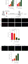
Tan IIA resulted in the repression of circulating Ang2 levels. (a) The Ang1 levels in the serum of mice treated with 0.3% CMC-Na and Tan IIA at day 21 postsurgery (n = 8). (b) The Ang2 levels in the serum of mice treated with 0.3% CMC-Na and Tan IIA at day 21 postsurgery (n = 8). (c) The Ang1 levels in the supernatant of HUVECs at 6 h, 12 h, and 24 h after DMSO or Tan IIA treatments (n = 3). (d) The Ang2 levels in the supernatant of HUVECs at 6 h, 12 h, and 24 h after DMSO or Tan IIA treatments (n = 3). The data were presented as mean ± SD. p < 0.05, ∗∗p < 0.01, ∗∗∗p < 0.001 (versus model group).
Figure 4
Figure 4
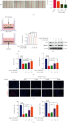
Tan IIA decreased EC permeability via inhibiting Ang2-mediated signaling cascade. (a) The HUVECs were treated with various concentrations of Tan IIA (0, 2.5, 5, and 10 μM) for 24 h. The area covered by migrating HUVECs was photographed by a phase-contrast microscopy. The migration of HUVECs was assessed on the basis of the wound closure area (n = 3). (b) Permeability measured in DMSO or Tan IIA treated HUVECs in the absence or presence of Ang2. The HUVEC permeability was quantified by the fluorescence of FITC-dextran (40 kD) collected in the bottom chamber (n = 3). (c) The expression of ZO-1 and Claudin-1 in the lysates of HUVEC treated with various concentrations of Tan IIA with or without the stimulation of 200 ng/ml of Ang2. GAPDH was used as a loading control. (d) Changes in the levels of ZO-1 and Claudin-1 were measured as pixel density and normalized to GAPDH (n = 3). (e) Representative immunofluorescence images of ZO-1 (green) and Claudin-1 (red) in HUVECs treated various concentrations of Tan IIA in the absence or presence of 200 ng/ml of Ang2. (f) Quantification of ZO-1 and Claudin-1 expression (n = 3). The data were presented as mean ± SD. p < 0.05, ∗∗p < 0.01, ∗∗∗p < 0.001 (versus control).
Figure 5
Figure 5
Tan IIA rescued the disrupted Tie2 activation mediated by Ang2. (a) Phosphorylation of Tie2 in HUVEC lysates 24 h after the treatment of 10 μM Tan IIA. GAPDH was used as a loading control (n = 3). (b) Representative immunofluorescence images of phospho-Tie2 (green) in HUVECs treated with various concentrations of Tan IIA in the absence or presence of 200 ng/ml of Ang2 (n = 3). (c) Phosphorylation of AKT, MLC, and total AKT, MLC in HUVEC lysates 24 h after the treatment of Tan IIA in the absence or presence of Ang2. GAPDH was used as a loading control. (d) Densitometric ratio for AKT activity was quantified. (e) Densitometric ratio for MLC activity was quantified (n = 3). The data were presented as mean ± SD. p < 0.05, ∗∗p < 0.01, ∗∗∗p < 0.001 (versus control).
Figure 6
Figure 6
Tan IIA potentiated the activation of Tie2 signaling pathway in vivo. (a) The mRNA expressions of indicated genes in the gastrocnemius muscle tissues were measured by real-time PCR (n = 8). (b) The mRNA expression of indicated genes in the transplanted HT-29 tumors was measured by real-time PCR (n = 8). (c) Phosphorylation of Tie2, AKT, MLC, and total Tie2, AKT, and MLC in the lysates from the gastrocnemius muscle tissues treated with various concentrations of Tan IIA. GAPDH was used as a loading control. (d) Phosphorylation of Tie2, AKT, and MLC and total Tie2, AKT, and MLC in the lysates from the harvested HT-29 tumors treated with various concentrations of Tan IIA. GAPDH was used as a loading control. (e) Densitometric ratios for the activities of Tie2, AKT, and MLC in the lysates from the gastrocnemius muscle tissues were quantified (n = 3). (f) Densitometric ratios for the activities of Tie2, AKT, and MLC in the lysates from the harvested HT-29 tumors were quantified (n = 3). The data were presented as mean ± SD. p < 0.05, ∗∗p < 0.01, ∗∗∗p < 0.001 (versus model group).
Figure 7
Figure 7
Tan IIA normalizes vessels in tumors and ischemic injury via regulating the Ang2/Tie2 signaling pathway. It revealed that Tan IIA maintained vascular stability by targeting the Ang2-Tie2-AKT-MLCK cascade.

Similar articles

Cited by

References

    1. Viallard C., Larrivee B. Tumor angiogenesis and vascular normalization: alternative therapeutic targets. Angiogenesis . 2017;20(4):409–426. doi: 10.1007/s10456-017-9562-9. - DOI - PubMed
    1. Zhao Y., Adjei A. A. Targeting angiogenesis in cancer therapy: moving beyond vascular endothelial growth factor. The Oncologist . 2015;20(6):660–673. doi: 10.1634/theoncologist.2014-0465. - DOI - PMC - PubMed
    1. Stylianopoulos T., Munn L. L., Jain R. K. Reengineering the tumor vasculature: improving drug delivery and efficacy. Trends in Cancer . 2018;4(4):258–259. doi: 10.1016/j.trecan.2018.02.010. - DOI - PMC - PubMed
    1. Dragneva G., Korpisalo P., Yla-Herttuala S. Promoting blood vessel growth in ischemic diseases: challenges in translating preclinical potential into clinical success. Disease Models & Mechanisms . 2013;6(2):312–322. - PMC - PubMed
    1. Li F., Sawada J., Komatsu M. R-Ras-Akt axis induces endothelial lumenogenesis and regulates the patency of regenerating vasculature. Nature Communications . 2017;8(1) doi: 10.1038/s41467-017-01865-x. - DOI - PMC - PubMed

MeSH terms

LinkOut - more resources